Poorly controlled diabetes may predispose patients to foot and lower leg ulceration, which, in severe cases, may progress to limb amputation (Grennan, 2019). Diabetic foot ulcers (DFUs) are complex, chronic wounds characterised by disrupted healing process and prolonged low-grade inflammation, reduced angiogenesis and defective extracellular matrix remodelling (Chang and Nguyen, 2021). Epidemiological studies indicate that these ulcers affect an estimated 25% of individuals with diabetes in their lifetime. The pathogenesis of DFUs is multifactorial, most commonly involving the interplay between peripheral neuropathy and vascular complications, such as peripheral artery disease (PAD), and repetitive mechanical trauma (Noor et al, 2017). Neuropathy causes sensory loss and foot deformities, leading to abnormal pressure and callus formation, which increases mechanical stress and, eventually, causes skin ulceration (Schaper et al, 2024). PAD impairs healing by reducing oxygen and nutrient delivery, leading to tissue ischaemia and raising infection risk.
Untreated hyperglycaemia further exacerbates wound chronicity through oxidative stress, endothelial dysfunction, and impaired cellular immune system, which collectively increase susceptibility to infection. Once infection occurs, the combination of ischaemia and immune dysfunction markedly elevates the risk of deep tissue involvement, osteomyelitis, and the need for surgical intervention, including amputation. (Grennan, 2019).
Recently, two standardised nature-derived protein fractions have been developed, DLBS1033, derived from the earthworm (Lumbricus rubellus), and striatin, derived from the snakehead fish (Channa striata; Dexa Laboratories of Biomolecular Sciences [DLBS], 2025). DLBS1033 contains lumbrokinases that improve systemic circulation through its antithrombotic and thrombolytic properties (Trisina et al, 2011). It has been shown to have a beneficial effect in ischaemic cases, including PAD and ischaemic stroke (Ndraha et al, 2013; Setyopranoto et al, 2016, Pinzon et al, 2021).
Currently, in some Southeast Asian countries, DLBS1033 has been used in clinical practice in the management of thrombosis-related diseases, such as ischaemic stroke, PAD, deep vein thrombosis and myocardial infarction (DLBS, 2025). In vitro and animal studies of striatin have shown its positive impact on cell proliferation, wound healing acceleration, and albumin level enhancement (Rahayu et al, 2016). In Indonesia, Cambodia and the Philippines, striatin has also been marketed and used as supplementary protein nutrition, particularly to accelerate wound healing (DLBS, 2025). However, there is limited clinical evidence regarding the combined use of these agents. The beneficial effects of DLBS1033 on vascular tissues and striatin on tissue repair, suggest there is potential for combining both agents to treat non-healing DFUs in patients with PAD.
The three reported cases indicate a valuable therapeutic potential of a combination of DLBS1033 and striatin in promoting the healing of non-healing DFUs complicated with suspected PAD.
Ethics
This case series was reviewed in accordance with institutional requirements. Formal ethics approval was not required for anonymised case reporting, but written informed consent for publication was obtained from the patients, and all identifying information was removed to protect confidentiality.
Case presentation
Case one
A 34-year-old man developed an infected, non-healing wound on his left foot over 2 weeks. He had a known history of uncontrolled type 2 diabetes (T2D) and was an occasional smoker, with no known history of hypertension, asthma or allergies.
Diagnostic, laboratory and wound assessments were performed on the day of consultation. His ABI values were 0.90 (left) and 0.88 (right). He also reported claudication, which, together with the ABI values, suggested mild bilateral PAD. Further examination, such as duplex ultrasonography, imaging or angiography, was not performed, due to the patient’s foot condition. He had elevated blood glucose levels, with fasting blood glucose (FBS) of 8.52 mmol/l, random blood sugar (RBS) of 14.1 mmol/l, and HbA1c of 77.0 mmol/mol. His creatinine level was normal at 70 µmol/l.
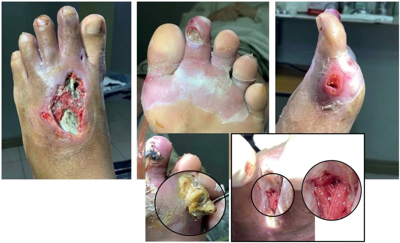
Key wound characteristics included the development of cellulitis, the presence of noticeable necrotic tissue and slough, and the secondary development of wound tunnels due to infection and swelling (Figure 1). Wound cultures were obtained through a sterile swabbing technique, appropriate aseptic precautions and standardised collection procedures were performed to minimise contamination and ensure that representative samples from necessary areas of the wound were adequately collected for culture analysis. The culture showed a positive infection, with a light growth of Proteus mirabilis after 72 hours of incubation. The wound was moderately infected according to the International Working Group on the Diabetic Foot/Infectious Diseases Society of America (IWGDF/IDSA) criteria, with a PEDIS (perfusion, extent, depth, infection, and sensation) grade 3. Based on the Wound, Ischemia, and Foot Infection (WIFI) classification system, the wound was classified as a grade 2 ulcer.
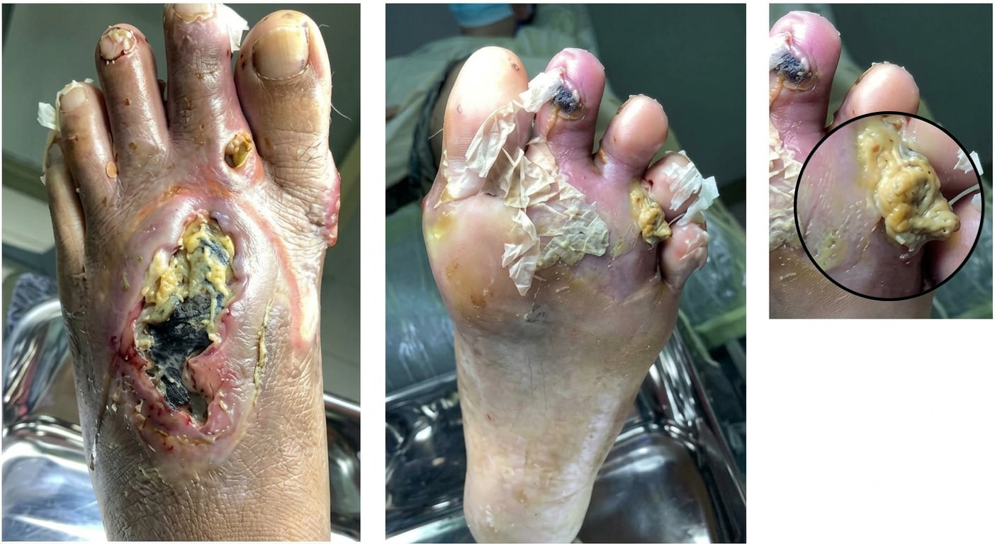
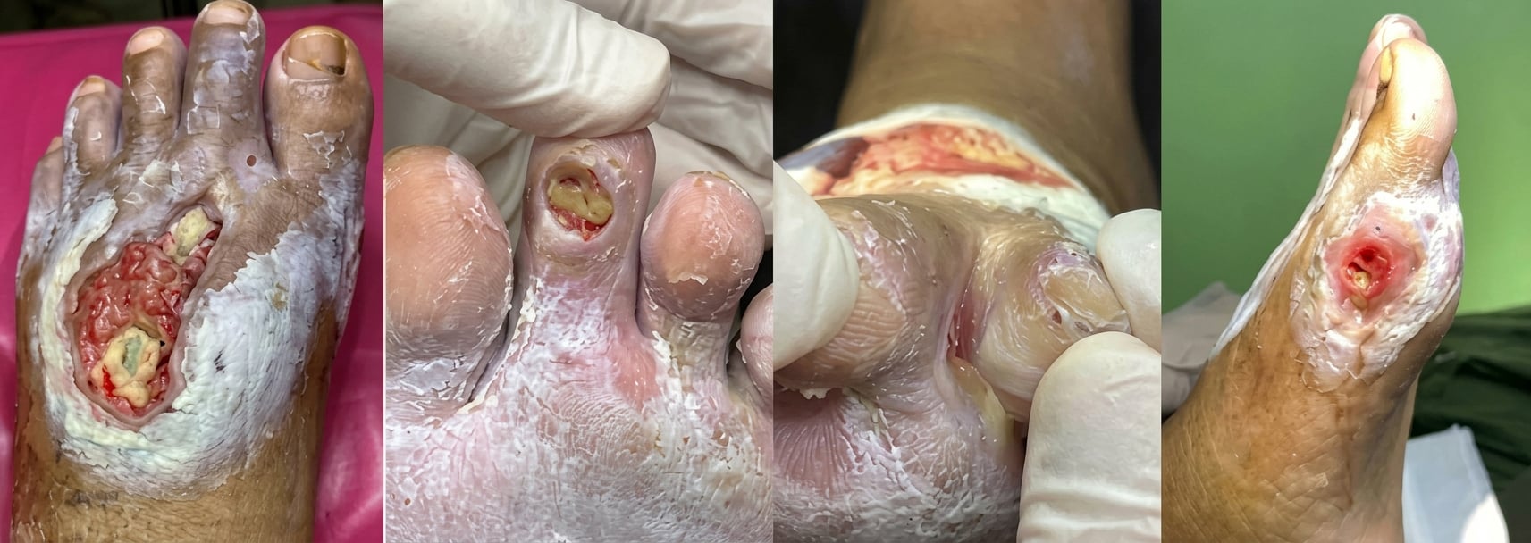
Sharp debridement was performed both manually and using an ultrasonic debridement machine. Post-debridement, vascular areas of the wound were exposed, resulting in more evident wound tunnels (Figure 2).
The following wound care techniques were sequentially performed post-debridement:
1. Spraying the wound with a wound cleanser.
2. Applying silver dressing directly to the wounds.
3. Applying zinc oxide ointment as a moisture barrier to the periwound area.
4. After wound cleansing and appropriate debridement, a silver hydrogel dressing was applied as the primary wound-contact dressing as clinically indicated.
5. The wound was then covered with a sterile, non-adherent absorbent as secondary cover to protect the wound bed, support exudate management, and minimise trauma during dressing removal.
Dressing changes were individualised according to wound assessment, exudate level, dressing integrity, and clinical signs of infection or deterioration. Wound cleansers are essential to standard wound care. The IWGDF guidelines emphasise the importance of therapeutic wound cleansing to prevent and manage wound infections (Senneville et al, 2023; Schaper et al, 2024). The wound cleanser contained hypochlorous acid and sodium hypochlorite as its active antimicrobial components. Throughout the first week post-debridement, wound care was performed every other day, and then every 3 days until the wound fully healed.
He was started on 490 mg DLBS1033 three times daily for PAD and 250 mg striatin twice daily to promote the wound healing process. Due to suspected PAD, rosuvastatin 20 mg once daily was also prescribed. For glycaemic control, he received sitagliptin 50 mg and metformin 500 mg fixed-dose combination twice daily, metformin 500 mg once daily, and glimepiride 2 mg once daily before breakfast. Additionally, to address the infection, he received ciprofloxacin 500 mg twice a day for 14 days. Ciprofloxacin was selected based on microbiological culture sensitivity results and its primary oral option in our clinical setting, bioavailability, and coverage against the identified organism. The dose of 500 mg twice daily for 14 days followed commonly utilised regimens for moderate diabetic foot soft tissue infection.
Clinical assessment on the patient’s wound did not raise sufficient suspicion to support involvement of deeper structures or bone at the time of evaluation. As such, there was no clinical justification for routine radiography for osteomyelitis assessment in this case. The patient was monitored clinically, and further diagnostic imaging would have been considered if findings suggestive of osteomyelitis or deeper infection had emerged. Nevertheless, he underwent careful clinical assessment for osteomyelitis, and no overt clinical findings strongly suggestive of advanced bone involvement were identified.
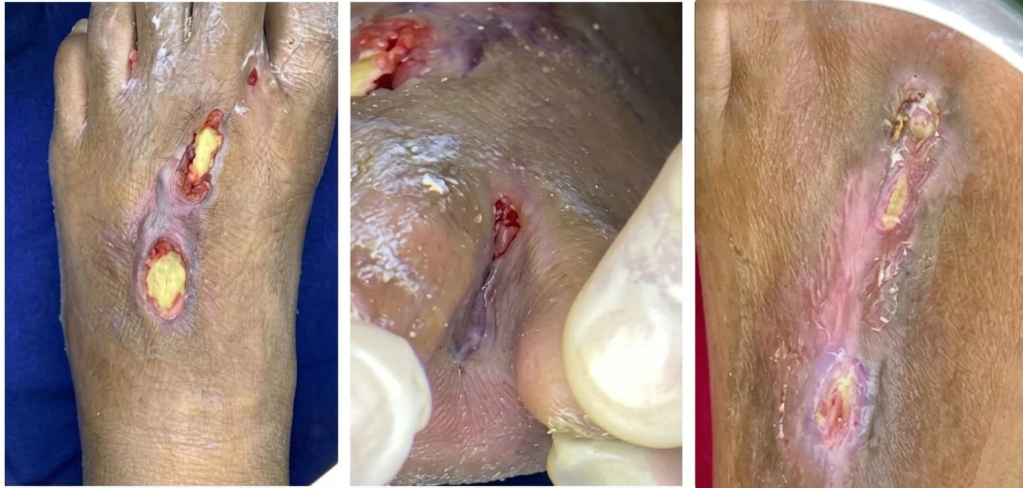
Seven days post-debridement, the wound had reduced in size and depth. The wound tunnels were closing with a granulation tissue formation (Figure 3). Progressive healing continued steadily, and by day 30, the tunnels had fully closed and scar tissue formed (Figure 4).
DLBS1033, a statin and glycaemic control medication for the management of the patient’s vascular disease and T2D were continued. Striatin was discontinued 3 months after the wound was completely healed. During observation and maintenance therapy with DLBS1033 and striatin, no adverse events were reported by the patient or physician.
Case two
An 82-year-old woman with uncontrolled T2D and controlled hypertension, and without any known drug or food allergies, developed a chronic, progressively enlarging, infected, non-healing wound on her right lateral malleolus and dorsum. The wound had persisted for 8 months. She was a former smoker with a history of 50 pack years and had been stopped for 12 years at presentation.
At consultation, she reported claudication. Such a clinical symptom and ABPI of 0.88 (left) and 0.86 (right) are suggestive of bilateral PAD. However, no further vascular examination, such as arterial Doppler scan, was performed, due to accessibility constraint in the study setting. Therefore, the PAD remained suspected. Her blood glucose parameters were above the normal ranges, with FBS 7.8 mmol/l, RBS 10.7 mmol/l and HbA1c 65.0 mmol/mol. Her creatinine level was normal at 86 µmol/l.
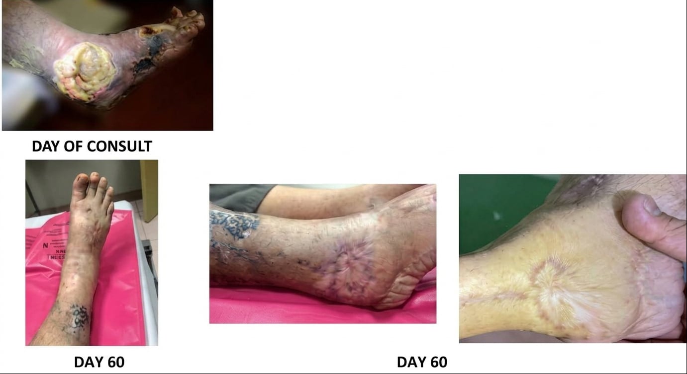
The wound was characterised by an exposed lateral malleolus with severe cellulitis and a necrotic area on the dorsum (Figure 5A). The specimen was obtained through wound swab culture following wound cleansing and debridement using aseptic technique. The swab was collected from viable wound tissue rather than superficial exudate. Although deep tissue culture is often preferred, appropriately performed wound swab cultures remain an accepted microbiological sampling method in diabetic foot infections and have demonstrated reasonable correlation with tissue cultures when standardised collection techniques (Levine) are observed. The wound culture findings were interpreted in correlation with the overall clinical presentation and infection severity. A light growth of Enterobacter cloacae was detected on wound culture after 72 hours of incubation. Based on the IWGDF/IDSA criteria, the severity of the infection was classified as a moderate infection (PEDIS grade 3) with a WIfI grade 3 ulcer.

Sharp debridement was performed manually and using an ultrasonic device. Five-step wound care techniques were performed, consistent with case 1. Dressings were changed every other day. On day 14 post-debridement, the silver dressing was replaced with a hydrofiber dressing to manage the exudate and maintain a moist healing wound environment. The hydrofiber dressing was utilised for extended wear, with dressing replacement individualised according to serial wound assessment, exudate level, and dressing integrity, generally within 7–10 days. Hydrofiber dressing was used based on its high absorptive capacity, moisture-balancing properties, and ability to conform closely to the wound bed while maintaining an optimal moist wound-healing environment. Hydrofiber dressings have been reported to assist in exudate management, reduce peri-wound maceration, and support autolytic wound healing (Whiteley, 2023). The wound was then covered with a sterile non-adherent absorbent for a secondary protective cover to protect the wound bed, support exudate management, and minimise trauma during dressing removal.
The patient was initiated on linagliptin 5 mg/empagliflozin 25 mg fixed-dose combination once daily for diabetes management, and rosuvastatin 20 mg once daily for PAD. For infection control, she received a 7-day course of ciprofloxacin 500 mg twice daily and clindamycin 300 mg every 6 hours. DLBS1033 490 mg three times daily and Striatin 250 mg twice daily were administered to help improve blood flow and wound healing. Ciprofloxacin was selected based on susceptibility of the isolated E cloacae and to provide Gram-negative coverage. Clindamycin was added for additional Gram-positive and anaerobic soft tissue coverage because of the associated cellulitis and necrotic wound involvement. The dosages utilised are commonly used oral regimens for moderate diabetic foot soft tissue infections. Duration of therapy was guided by serial clinical assessment and response to treatment.
On day 21 post-debridement, there was significant granulation tissue formation, reduced wound size and depth, and decreased inflammation (Figure 5B). On day 40 post-debridement, the wound exhibited a stable eschar, progressive granulation tissue formation, further size reduction, minimal swelling, absent signs of necrosis or gangrene, and the emergence of new epidermal tissue (Figure 5C). The wound had completely healed by day 60 post-debridement (Figure 5D). Figure 6 illustrates the overall wound healing progression over 60 days, compared to the initial presentation.
Maintenance treatment with DLBS1033 490 mg three times daily, high-intensity statin, and anti- hyperglycaemic agents was continued to manage the PAD and diabetes. Striatin 250 mg twice daily was discontinued 3 months after complete wound healing.
No adverse events were reported or observed during the treatment with DLBS1033 and striatin.
Case three
A 49-year-old woman presented with a non-healing and infected atypical diabetic foot ulcer on her left foot, which had been present for 2 months following self-treatment using an unspecified ointment she had purchased online. The lesion demonstrated clinically significant infection characterised by hypertrophic scar breakdown, underlying deep ulceration and surrounding inflammation.
She had uncontrolled T2D and a 15-pack-year smoking history, with no hypertension, asthma or known allergies.
Clinical examination strongly suspected bilateral PAD, with ABI values of 0.90 bilaterally, and claudication symptoms. Laboratory test showed high blood glucose levels with FBS 8.4 mmol/l, RBS 12.3 mmol/l and HbA1c 78.1 mmol/mol, and a normal creatinine level of 78 µmol/l.
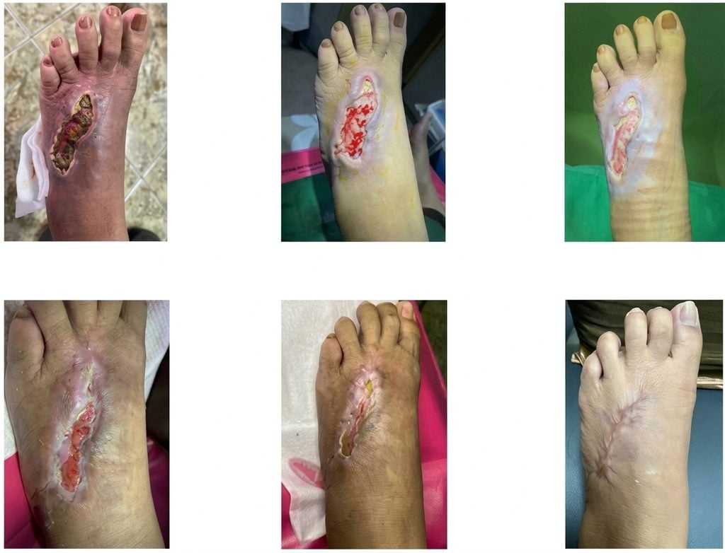
On the consultation day, the lesion demonstrated clinically significant infection characterised by hypertrophic scar breakdown, underlying deep ulceration, erythema and surrounding inflammation (Figure 7A). Similar with the other two cases, the wound specimens were obtained using a sterile swab technique following wound cleansing, irrigation, and debridement to minimise superficial contamination. Samples were collected from viable wound tissue and deeper affected areas while avoiding superficial exudate and necrotic debris. Specimens were submitted for aerobic culture and antibiotic susceptibility testing according to standard laboratory protocols. Although deep tissue culture is often preferred, properly obtained wound swab cultures remain an accepted microbiological sampling method when performed with adequate wound bed preparation and aseptic technique. Wound culture revealed light growth of Enterobacter agglomerans after 72 hours of incubation. According to the IWGDF/IDSA criteria, the wound was moderately infected (PEDIS grade 3) with a grade 3 ulcer under the WIfI classification.
A sharp debridement was performed manually and using an ultrasonic device. After debridement, vascular areas were exposed (Figure 7B). The similar standardised wound care protocol, as applied in previous cases, was performed. The wound care was scheduled every other day for the first week, then every 3 days until the wound healed. This practice is preferred when improving wound, such as predominance of granulation tissues, clean, with minimal exudate, is observed.
The patient was initiated on a fixed-dose combination of sitagliptin 50 mg/metformin 500 mg twice daily, metformin 500 mg once daily, glimepiride 2 mg once daily, to control her diabetes and rosuvastatin 20 mg once daily for her PAD. The patient was given the antibiotic coamoxiclav 625 mg three times daily for 10 days. The 10-day duration was guided by the clinical severity of the infection, associated cellulitis and serial wound assessment during follow-up. Treatment with DLBS1033 490 mg three times daily and striatin 250 mg twice daily was also started to promote wound healing.
Progressive healing was observed from day 1 to day 7 post-debridement, significantly reducing wound size and depth on day 18 (Figures 7C, 7D and 7E). The wound showed significant granulation tissue formation, with no signs of swelling, erythema, or necrosis compared to the initial presentation. On day 32, the wound appeared entirely healed (Figure 7F).
The patient and the physician reported or observed no adverse events during the treatment and maintenance therapy with DLBS1033 and striatin. Due to PAD and diabetes, maintenance treatment with DLBS1033 490 mg, high-intensity statin, and antidiabetic medications were continued. Three months following the complete wound healing, Striatin was discontinued.
Discussion
These three cases are among the first cases to explore the combination of DLBS1033 and striatin for non-healing DFUs in patients with T2D and suggestive PAD.
Non-healing wounds in diabetes with PAD or strongly suggested PAD are challenging, as the wounds become stalled in the inflammatory stage. The normal wound healing process involves several critical phases: inflammation, proliferation, and remodeling of the extracellular matrix that restores the dermis. Following an injury, neutrophils migrate to the wound area and secrete matrix metalloproteinase (MMP)-8 to debride the wound and cleave damaged collagen type I. By removing the dead, necrotic, or infected tissues, the wound debridement will promote the healing process. Neutrophils also secrete reactive oxygen species (ROS) to eradicate bacteria and control thrombus formation, or blood clots, to halt bleeding (Chang and Nguyen, 2021). Diabetes may impair neutrophil function, increasing the risk of infections, such as progressive and necrotic infections (Senneville et al, 2023). PAD further complicates healing due to narrowed blood vessels in the wound area, leading to hypoxia, which impairs oxygen-dependent processes such as ROS production (Chang and Nguyen, 2021).
A 12-month study by Ndosi et al (2018) showed that patients with clinically infected DFU on antibiotics or antimicrobial treatment presented only 44.5% healing incidence, 17.4% recorded amputation, revascularisation surgery of 6.0%, and a mortality rate of 15.1%. Furthermore, in PAD cases with non-healing wounds, high-dose statin therapy was administered due to the high likelihood of the underlying atherosclerotic condition. Statins provide cardiovascular risk reduction and pleiotropic vascular benefits, including endothelial stabilisation, anti-inflammatory effects, and improved perfusion, which may contribute to enhance wound healing and limb preservation (Oesterle et al, 2017; Chou et al, 2022). Statin therapy in PAD is already considered standard vascular risk management (ADA, 2026, Gornik et al, 2024, Mazzolai et al, 2024, Schaper et al, 2024, Senneville et al, 2023).
DLBS1033 has been studied for years for its antithrombotic and thrombolytic properties in both preclinical (Trisina et al, 2011), and clinical studies (Ndraha et al, 2013; Setyopranoto et al, 2016; Pinzon et al, 2021). Furthermore, DLBS1033 also possesses several pleiotropic activities supporting its main mechanism of action, such as the anti-inflammatory activity, smooth muscle cell viability and migration inhibition, and plaque stabilisation enhancement (Trisina et al, 2011). Its safety profile has been proven through toxicity studies (Sukandar et al, 2014; Sajuthi et al, 2011), and studies in healthy volunteers (Gayatri et al, 2018, Tjandrawinata et al, 2016). Due to its capacity to improve blood circulation, DLBS1033 treatment has been shown to have clinical benefit for patients with ischaemic diseases, such as PAD (Ndraha et al, 2013), and ischaemic stroke (Setyopranoto et al, 2016; Pinzon et al, 2021).
Striatin has been shown to promote cell growth during the healing process, mainly in the proliferation phase (Rahayu et al, 2016). This ability may be from its capacity to enhance fibroblast cell growth (Rahayu et al, 2016), which plays a crucial role in tissue repair (Addis et al, 2020).
The effect of striatin was further evaluated using a wound-induced animal model. The study revealed a significant reduction in the wound area, accompanied by a significant increase in serum albumin level compared to the control group, indicating an enhanced nutrition and a more favorable environment for tissue repair (Rahayu et al, 2016).
In each case in this report, a combination therapy with DLBS1033 (490 mg three times daily) and Striatin (250 mg twice daily) was administered for 30–60 days post-debridement. Our findings align with previous clinical studies of DLBS1033 in improving blood circulation (Trisina et al, 2011), and the role of striatin in promoting wound healing (Rahayu et al, 2016). These reports indicate the promising therapeutic potential of a combination therapy such as this in managing DFUs. All patients received the combination therapy throughout their observation period, lasting 30–60 days.
Overall, all cases demonstrated satisfactory wound healing, with no revascularisation or amputation required. Case one showed rapid regression of wound size and depth, progressive tissue granulation and closure of wound tunnels. The new scar formation indicated that the wound was already in the remodelling phase. In the second case, the wound totally healed. Case three reached the remodelling phase. In all three cases, the use of DLBS1033 490 mg three times daily was even maintained for longer periods as a part of the patients’ vascular-related therapy, alongside with statin and anti-diabetes medications; while striatin was discontinued 3 months after the wound was completely healed.
Limitations
Despite the promising findings reported in these three patients, we acknowledge that these are case reports, not a clinical study.
There were several limitations in our current cases. First, we noticed that ABI alone is insufficient to reliably determine the severity of PAD in a person with diabetes or exclude clinically significant ischaemia, particularly because arterial calcification may affect ABI interpretation.
Second, according to current diabetic foot and PAD guidance, suspected PAD in a person with diabetes and a foot ulcer, particularly in the presence of claudication symptoms, should prompt further vascular assessment where available, such as pedal arterial duplex ultrasound, toe pressure/TBI, or referral to a vascular specialist (Gornik et al, 2024; Mazzolai et al, 2024). The absence of these advanced vascular investigations represents a major limitation of the case series and should not be interpreted as a recommendation or acceptable standard for routine clinical practice. In fact, in routine practice, suspected PAD in this context should prompt further vascular assessment and/or vascular specialist referral where clinically indicated. Therefore, we carefully selected the term “suspected PAD” rather than “confirmed PAD” for the case series, with PAD severity was preferably not to be specifically mentioned.
Conclusion
The three reported cases suggest a therapeutic potential of the combination of DLBS1033 and striatin in promoting wound healing in DFUs. No adverse events were reported by the patients or observed by the physician. These early findings suggest that this therapeutic approach holds a promise for improving outcomes in diabetic patients with non-healing DFUs suspected with PAD. However, further randomised clinical trials are needed to provide a more robust evidence for the use of this new combination therapy in DFUs treated in clinical practice.n