

Impaired beta-cell function is a defining feature of diabetes pathophysiology, with progressive loss of insulin-secreting capacity. Decline is typically faster and more severe in type 1 diabetes, driven by autoimmune destruction, versus the slower, more progressive loss that occurs in type 2 diabetes, often against a background of insulin resistance (Maddaloni et al, 2022; ADA Professional Practice Committee, 2026; Vinay et al, 2026). Intermediate patterns occur in immune-mediated, pancreatogenic and monogenic diabetes.
Diabetes type is not always clear at presentation, but accurate classification is essential as it guides treatment, medication choice, education and use of diabetes technologies. The principal factor driving differences in treatment between type 1 and type 2 diabetes is severe insulin deficiency. Individuals with absent or near-absent endogenous insulin secretion require treatment approaches aligned with type 1 diabetes, regardless of the underlying cause of beta-cell failure.
Classifying diabetes correctly
Features pointing to type 1 diabetes include the following (ADA Professional Practice Committee, 2026):
- Younger age (<35 years).
- Lower BMI (<25 kg/m2).
- Rapid onset of osmotic symptoms.
- Unintentional weight loss.
- Plasma glucose >20 mmol/L.
- Sometimes, diabetic ketoacidosis (DKA) at presentation.
Anyone with symptoms suggestive of type 1 diabetes should be seen face-to-face on the same day as an urgent matter, with fingerprick glucose and ketone testing included, as DKA can deteriorate rapidly and be life-threatening (John, 2011; NICE, 2022). Indeed, the Care Quality Commission (2026) recommends that a blood glucose monitor, test strips and lancets should be immediately available in general practice to assess blood glucose levels.
If there is high suspicion of type 1 diabetes, refer immediately to hospital/specialist services for insulin initiation. Autoantibody testing can follow later if deemed necessary.
Autoantibody testing
The autoantibodies implicated in type 1 diabetes are known as islet cell antibodies (ICAs). These include antibodies against GAD, IA2, insulin and ZnT8, and are used alongside clinical assessment to help confirm type 1 diabetes (Jamjoom et al, 2024).
Almost 90% of people newly diagnosed with type 1 diabetes have at least one detectable autoantibody; around 53% test positive for GAD alone, 83% for GAD and/or IA2, and about 88% for one or more of GAD, IA2, ZnT8 and/or insulin (Carlsson et al, 2020).
The American Diabetes Association (ADA) and European Association for the Study of Diabetes (EASD) joint consensus on type 1 diabetes does not recommend measuring ICAs unless there are clear features suggesting type 1 diabetes (Holt et al, 2026), as false-positives can occur. Where there is only one positive antibody, interpret with caution where type 1 diabetes is unlikely based on clinical signs.
Staging of type 1 diabetes
Type 1 diabetes is now recognised as a progressive autoimmune condition with three distinct stages, which starts long before symptoms appear. Pre-symptomatic type 1 diabetes (stages 1–2) can be detected via autoantibody screening, although no UK national programme yet exists. Screening aims to identify people before substantial beta-cell loss has occurred, to reduce rates of DKA at diagnosis and to identify those who may benefit from disease-modifying immunotherapy that can delay progression to stage 3 (symptomatic) type 1 diabetes (Leichter et al, 2025).
This staging incorporates what was formerly called latent autoimmune diabetes in adults (LADA), which is now considered a subset of adult-onset autoimmune diabetes rather than a separate condition. Most people with this subtype test positive for at least one islet autoantibody (usually GAD) but progress to insulin dependence more slowly than those with classic type 1 diabetes, so it is often misdiagnosed as type 2 diabetes (Jamjoom et al, 2024). Misdiagnosis risks delayed insulin initiation, inappropriate treatment with non-insulin therapies, and metabolic decompensation, including DKA.
MODY
Maturity-onset diabetes of the young (MODY) is a monogenic form of diabetes caused by a single-gene mutation affecting beta-cell function. Core features include young onset (before age 35 years), strong family history and negative autoantibodies. Some insulin production is retained compared with type 1 diabetes.
Many people with MODY are misdiagnosed with type 1 diabetes and started on insulin unnecessarily, although some subtypes (especially HNF1A) respond well to sulfonylureas instead.
C-peptide may help distinguish MODY from longer-duration type 1 diabetes (Leighton et al, 2017). The MODY Probability Calculator, available at https://www.diabetesgenes.org, can help identify who should be tested for monogenic diabetes (University of Exeter, 2026).
What is C-peptide?
Insulin and C-peptide are co-released from proinsulin in equimolar amounts (Jones and Hattersley, 2013). Endogenous insulin is hard to measure directly because of its short half-life (around 5 minutes) and rapid fluctuation (Polonsky and Rubenstein, 1984; Lin et al, 2025). Up to 80% of insulin is also extracted by the liver on first pass, so peripheral levels underestimate true pancreatic output (Najjar and Perdomo, 2019). In contrast, C-peptide has a longer half-life (30–35 minutes) and minimal hepatic extraction, so its peripheral concentration more accurately reflects endogenous insulin production (Polonsky and Rubenstein, 1984).
C-peptide may be used as a surrogate marker of beta-cell function when assessing insulin secretory capacity, but multiple factors can influence results. It is not a routine investigation in primary care, and it should be reserved for situations where diagnostic uncertainty persists.
C-peptide and autoantibody tests give complementary information; antibodies show whether the immune system is attacking beta-cells, while C-peptide shows remaining beta-cell function:
- Early autoimmune diabetes has positive antibodies with preserved C-peptide.
- Later stages show persistent antibodies with low or absent C-peptide.
- Very late stages may become seronegative despite complete C-peptide loss, with rare seronegative type 1 diabetes also recognised (ADA Professional Practice Committee, 2026).
Using C-peptide in clinical practice
Because C-peptide reflects endogenous beta-cell activity, it can help classify diabetes type in selected clinical contexts. It may also help predict diabetes-related vascular complications, guide pharmacotherapy choices and evaluate hypoglycaemia (Vinay et al, 2026).
Distinguishing diabetes type
C-peptide may help differentiate diabetes type where there is doubt – for example, in an adult with atypical features at or after diagnosis (higher BMI, slow progression, strong family history or features suggestive of MODY/LADA) – by distinguishing insulin deficiency (likely in type 1 diabetes) from preserved endogenous insulin (more consistent with type 2 diabetes or MODY).
Low or undetectable C-peptide levels support a type 1 diabetes or autoimmune aetiology, whereas elevated or normal levels suggest type 2 diabetes with intact or excessive beta-cell output (e.g. insulin resistance). Note, however, that in the first 1–3 years after type 1 diabetes diagnosis, there may still be residual beta-cell function, so C-peptide may still be detectable or even near normal (and potentially misread as indicating type 2 diabetes or MODY). Additionally, around the time of diagnosis, DKA and glucotoxicity can temporarily suppress beta-cell function, so a low C-peptide may not reliably reflect true beta-cell reserve (recovery can occur once glucose levels improve).
By 3 years post-diagnosis, beta-cell destruction is typically near complete and C-peptide levels are very low (<200 pmol/L) or undetectable. Thus, negative antibodies with preserved C-peptide after 3 years should prompt consideration of MODY or type 2 diabetes, while positive antibodies with low C-peptide suggest autoimmune diabetes.
At type 1 diabetes presentation, 80–90% of beta-cell loss has typically occurred, although a “honeymoon phase” may bring temporary recovery (lasting weeks to months) with rising C-peptide. For most people, ongoing autoimmune destruction causes C-peptide to decline to very low or undetectable levels within a few years.
C-peptide typically declines gradually in type 1 diabetes: DCCT data showed 48% of participants retained some beta-cell function at 5 years, falling to 8% by 5–15 years’ duration (DCCT Research Group, 1987). Younger age of type 1 diabetes onset (<10 years) may be linked to a faster decline (Kuhtreiber et al, 2015).
Re-evaluating a classification of type 1 diabetes after several years
In those with a long-standing type 1 diabetes diagnosis but a clinical picture suggesting possible type 2 diabetes or MODY (e.g. low insulin requirements, obesity, strong family history, metabolic syndrome), C-peptide may be checked before deintensifying or withdrawing insulin.
Detectable or robust C-peptide years after diagnosis suggests misclassification and should prompt a review of diagnosis and treatment.
What do the guidelines recommend?
Most guidelines recommend against measuring C-peptide if diabetes duration is under 3 years, although it can be measured earlier to aid diagnosis of insulin deficiency, if insulin withdrawal is being considered, or if a monogenic form of diabetes is suspected.
According to NICE (2026), type 1 diabetes should be considered in an adult presenting with hyperglycaemia (random plasma glucose >11 mmol/L) plus, typically, one or more of the following:
- Ketosis – ketones are high if above 2+ in the urine or above 3 mmol/L in the blood.
- Rapid weight loss.
- Age of onset younger than 50 years.
- BMI below 25 kg/m2.
- Personal and/or family history of autoimmune disease.
- Do not rule out a diagnosis of type 1 diabetes if an adult has a BMI of 25 kg/m2 or above, or is aged 50 years or over; use clinical judgement.
- Assess for underlying causes such as pancreatic cancer if aged ≥60 years with weight loss and new-onset diabetes.
- Measure diabetes-specific autoantibodies in adults with an initial type 1 diabetes diagnosis; false-negative rates are lowest at diagnosis and reduced by testing two different autoantibodies (at least one positive).
NICE recognises C-peptide as a marker of endogenous insulin – plasma C-peptide <200 pmol/L with a paired glucose >4 mmol/L usually indicates severe insulin deficiency – but does not recommend its routine use to confirm type 1 diabetes, reserving it for cases where clinical features suggest type 1 diabetes but autoantibodies are negative.
The ADA supports C-peptide testing mainly as a diagnostic tool to distinguish diabetes types, and provides a useful flowchart for investigating suspected type 1 diabetes (ADA Professional Practice Committee, 2026).
Utility beyond clarifying diabetes type
C-peptide testing also has clinical utility beyond classification, offering insight into beta-cell function, treatment needs and metabolic risk, although these uses are not yet routine in everyday practice. Its potential role in type 2 diabetes management is an emerging area of interest (Lin et al, 2025).
Predicting the need for insulin
Individuals with absolute insulin deficiency (C-peptide <200 pmol/L after mixed-meal stimulation, or fasting C-peptide <80 pmol/L) will need insulin regardless of diabetes type (Vinay et al, 2026). It may also help identify beta-cell exhaustion, and thus requirement for insulin therapy, in long-standing type 2 diabetes (Leighton et al, 2017), although evidence for its predictive value remains limited (Vinay et al, 2026).
Guiding treatment decisions
There may be a role in predicting treatment response to non-insulin therapies in type 2 diabetes, although evidence is limited and inconsistent (Lin et al, 2025). Very low C-peptide almost always indicates a need for insulin, while preserved secretion suggests the individual may respond well to GLP-1 receptor agonists, SGLT2 inhibitors or other non-insulin therapies. It can also help to determine whether insulin can safely be reduced or stopped (ADA Professional Practice Committee, 2026).
Evaluating hypoglycaemia
C-peptide differentiates endogenous from exogenous insulin. In cases of hypoglycaemia, high insulin with high C-peptide suggests endogenous hyperinsulinism (for example, due to insulinoma or treatment with a sulfonylurea), while high insulin with low C-peptide strongly suggests the hypoglycaemia is due to exogenous insulin administration (Vinay et al, 2026).
Predicting diabetes complications
An emerging area of research is whether C-peptide can help predict diabetes complications, with lower concentrations linked to poorer glycaemic control, higher HbA1c and, potentially, increased complication risk (Leighton et al, 2017).
How is C-peptide measured?
There are several ways to measure C-peptide, using blood or urine samples and either fasted or non-fasted. The ADA/EASD consensus recommends to measure non-fasting C-peptide (within 5 hours of a carbohydrate-containing meal) alongside a concurrent glucose concentration; if the glucose level is <4 mmol/L and C-peptide <600 pmol/L, the test should be repeated (Holt et al, 2026).
However, there is no definitive guideline on the preferred method within clinical practice settings; check with your local laboratory regarding which tests are available in your area and the preparation required.
Avoid testing in the following scenarios:
- Within 2 weeks of a hyperglycaemic emergency (e.g. DKA or hyperosmolar hyperglycaemic state), as C-peptide may be transiently suppressed.
- If significant hypoglycaemia in the previous 12 hours.
Repeat the test if results are inconsistent with the clinical picture.
Further information is available at:
- https://www.diabetesgenes.org
- https://www.exeterlaboratory.com/test/c-peptide-plasma/
- NHS Tayside Diabetes Managed Clinical Network: https://bit.ly/46Basps
Interpreting results
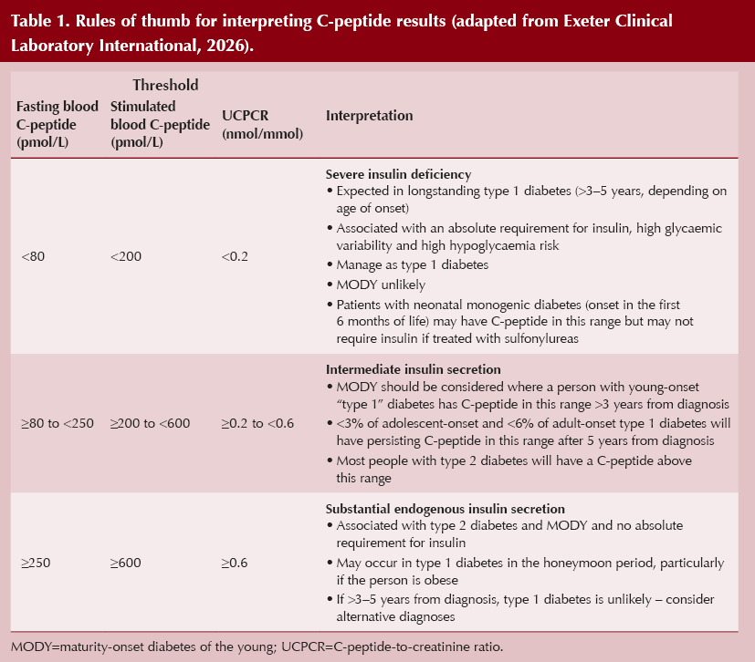
Rules of thumb for interpretation are summarised in Table 1 (Exeter Clinical Laboratory International, 2026). However, there are several situations that can affect C-peptide concentration independently of beta-cell reserve (Vinay et al, 2026). Practically, the most relevant issues are:
Renal failure: C-peptide will be falsely raised with more severe renal failure, as the kidneys are the primary clearance organ. Therefore, be cautious with high results if estimated glomerular filtration rate is <30 mL/min/1.73 m2.
Lack of stimulation: A glucose level ≥8 mmol/L is generally needed to adequately stimulate C-peptide production. Therefore, a sample drawn at low-normal glucose may appear falsely low.
- Where a random sample is used (and therefore there is risk that C-peptide is poorly stimulated), a repeat test should be considered for low results, especially if close to a clinically relevant threshold.
Acute hyperglycaemia: Glucotoxicity can temporarily suppress beta-cell function and give falsely low results that recover with improved control. Do not measure C-peptide during, or in the 2 weeks following, a hyperglycaemic emergency (diabetic ketoacidosis or hyperosmolar hyperglycaemic state).
Hypoglycaemia: Hypoglycaemia can transiently suppress endogenous insulin secretion and give misleadingly low results – avoid testing C-peptide within 12 hours of a hypoglycaemic episode.
Therefore, a C-peptide result should never be interpreted in isolation – it must be read alongside other clinical and biochemical findings, particularly when results are close to clinical thresholds.
Conclusion
C-peptide is a valuable tool when diabetes classification is uncertain, helping to assess endogenous insulin secretion and clarify insulin deficiency. It must always be interpreted in context, however, taking into consideration glucose levels at sampling, renal function, diabetes duration, autoantibody status, BMI and recent hyper- or hypoglycaemia. Used appropriately, it supports accurate diagnosis, guides treatment and can flag misclassification, but should never be used in isolation.
Acknowledgement
The author thanks Angus Jones for his feedback on the early draft of this article.




Resources from Diabetes UK to support earlier identification of type 1 diabetes.
12 Nov 2026