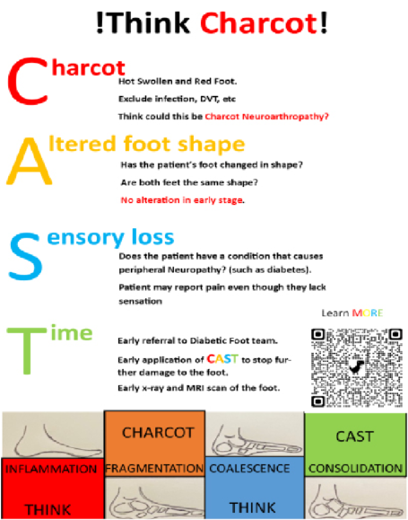
Charcot neuro-osteoarthropathy (CNO) is a rare, but severe, complication of neuropathy characterised by progressive bone and joint destruction that can lead to deformity, ulceration and limb loss. Early recognition is essential, as timely offloading during the acute inflammatory phase (stage 0) can prevent irreversible collapse. However, early-stage CNO frequently presents with subtle signs, such as erythema, swelling, warmth or mild discomfort despite normal radiographs, making diagnosis particularly challenging (Rajbhandari et al, 2002; Holmes et al, 2015).
Missed or delayed diagnosis remains common across healthcare settings in the NHS. Schmidt et al (2017) found that 67% of primary care and internal medicine practitioners had little or no knowledge of CNO. Sherratt et al (2024) report that 64% of patients are initially misdiagnosed in primary care, with a mean diagnostic delay of 81 days in those without a prior history of CNO. CNO is frequently mistaken for cellulitis, osteomyelitis, gout, deep vein thrombosis or simple soft-tissue injury – all of which are far more prevalent and, therefore, more readily considered by non-specialists (Safavi et al, 2022). Limited clinician education and awareness, poor diagnostic thresholds and variable referral pathways further contribute to delays.
The consequences of late recognition are substantial. In the UK, the cost of managing CNO is estimated at more than £6.5 million annually, with nearly half of affected feet ultimately requiring surgery and up to 8% progressing to amputation (Kerr et al, 2014; Metcalf et al, 2018; Gratwohl et al, 2022). Pinzur and Schiff (2017) demonstrated significantly higher success rates with earlier surgical intervention, yet national pathways fail to mandate the importance of early surgical referral.
Despite guidelines from NICE (2019) and International Working Group on the Diabetic Foot (IWGDF; Wukich et al, 2024) providing broad principles for assessment and management, ambiguity persists regarding referral to patient contact timelines, imaging standards and timeframes, and responsibility for specialist foot surgeon review. These gaps may contribute to variation in practice, loss of accountability and delayed escalation of diagnosis and thorough assessment and management.
This case-based review highlights how systemic ambiguities contributed to a delayed diagnosis of acute CNO in a patient who subsequently developed severe deformity requiring significant reconstructive surgery. The case is used to illustrate common diagnostic pitfalls and to propose clearer pathways to improve early recognition and specialist foot surgeon involvement within UK healthcare services.
Case presentation
A 63-year-old man with a history of type 2 diabetes presented to his GP with a several-day history of unexplained pain, erythema and swelling affecting his left foot. There was no history of trauma. Suspecting cellulitis, the GP commenced a 7-day course of flucloxacillin 500 mg four-times daily. At review following completion of antibiotics, the patient reported no improvement in symptoms. A plain radiograph of the foot was arranged, which appeared normal, and he was subsequently referred to the community podiatry service.
He was assessed 3 days later by the podiatry team. Infrared dermal thermometry demonstrated a 1.2°C temperature difference between the affected and contralateral foot. Peripheral neuropathy testing using a 10 g monofilament was documented as normal. Based on these findings, acute Charcot neuro-osteoarthropathy was not suspected and the patient was discharged with orthotic insoles.
Over the following 8 weeks, the patient experienced progressive worsening of pain, swelling and deformity. He re-attended his GP with marked midfoot collapse. An urgent referral was made to the regional diabetic foot clinic, where further clinical evaluation raised concern for acute Charcot neuro-osteoarthropathy. MRI of the left foot confirmed stage 3 Charcot neuroarthropathy, with evidence of significant osseous destruction and midfoot instability.
The patient was treated with immobilisation in a total contact cast until clinical remission of the acute phase was achieved. Despite this, he subsequently developed recurrent plantar midfoot ulceration related to progressive deformity. Owing to persistent instability and ulcer risk, he later underwent extensive reconstructive surgery performed by the podiatric surgery team. He remains under follow-up for long-term offloading and surveillance.
Discussion
Early CNO is notoriously difficult to recognise, particularly during the prodromal “stage 0” phase when clinical changes precede radiographic abnormalities. Patients commonly present with non-specific signs, such as erythema, warmth, oedema and mild discomfort, all of which overlap with several more prevalent differential diagnoses (Holmes et al, 2015; Schmidt, 2020). Radiographs are frequently normal at this stage, contributing to significant diagnostic uncertainty and false reassurance among non-specialist clinicians (Artz et al, 2023).
Traditional bedside assessments further complicate early identification. The Semmes-Weinstein 10 g monofilament test, widely used in primary and community settings to assess peripheral neuropathy, primarily detects large-fibre (A-beta) dysfunction. However, CNO may develop in the presence of small-fibre or patchy neuropathy despite preserved monofilament sensation (Burgess et al, 2021; Carmichael et al, 2021). Reported sensitivity of the monofilament test for neuropathy may be as low as 41%, leading to false-negative results in patients who are at risk of developing CNO (Dros et al, 2009).
Additionally, autonomic and motor neuropathy assessments are not endorsed by NICE or IWGDF in suspicion of CNO. This is particularly relevant, as early CNO frequently presents with autonomic dysregulation, characterised by increased perfusion, impaired vasomotor control, and associated hyperaemia, all mediated by small fibres and considered key early drivers of CNO pathophysiology that may precede overt sensory loss (Petrova and Edmonds, 2008; Kaynak et al, 2013). In the present case, preserved monofilament sensation contributed to a low index of suspicion and discharge from podiatry without offloading or escalation.
Binns-Hall et al (2018) demonstrated that combining a ‘DPN-Check’ assessment, quantifying large-fibre sural nerve conduction, with Sudoscan, which evaluates small-fibre function via electrochemical skin conductance, substantially increases diagnostic accuracy. When used alongside 10-g monofilament testing, this multimodal approach increases sensitivity for detecting early neuropathy to 94%. Such specialised diagnostic modalities could be utilised by specialist foot surgeons, and provide clinically meaningful insights that facilitate identification of stage 0 CNO. These findings reinforce the value of early referral to specialist foot surgeons to optimise early diagnostic accuracy and subsequent management.
Infrared dermal thermometry is frequently recommended as an adjunctive assessment tool; however, diagnostic thresholds vary widely across the literature. Armstrong and Lavery (1997) reported inter-limb temperature differences of 2.2–13.2°C in confirmed CNO, whereas Jones et al (2023) report in their systematic review differences as low as 0.7°C. External factors, such as rest duration and ambient temperature, further limit accuracy (Dallimore et al, 2020). Although a temperature difference of ≥2°C is commonly used as a pragmatic indicator of active disease, no CNO-specific validation studies have established its sensitivity or specificity. Consequently, mild temperature asymmetry – such as the 1.2°C difference observed in this case may be misinterpreted as insufficient to support a diagnosis, contributing to missed opportunities for early offloading or surgical referral.
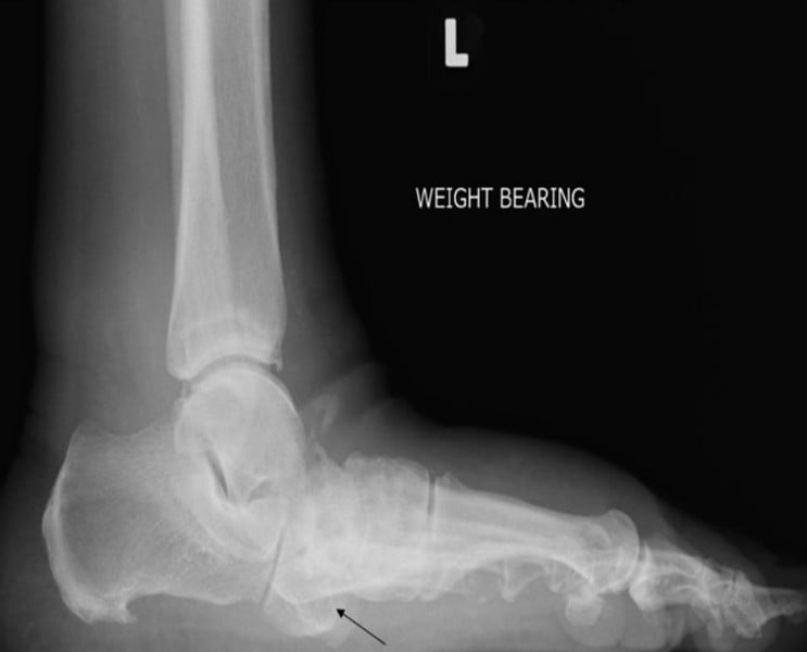
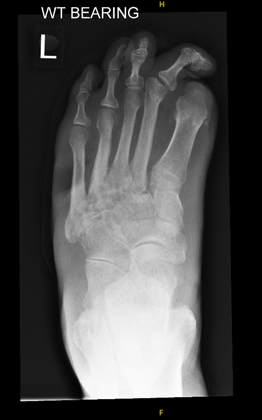
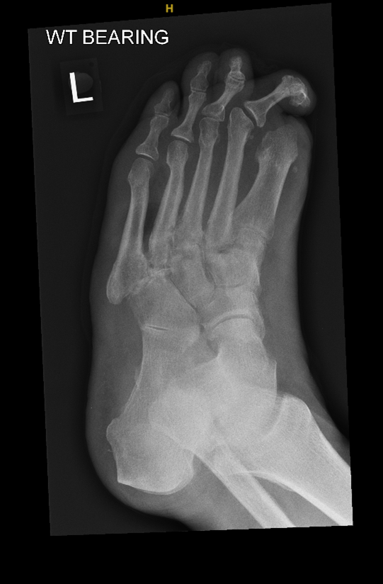
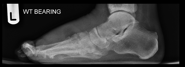
Plain radiography is widely accessible but offers limited diagnostic value in early CNO. Radiographic sensitivity in stage 0 is less than 50%, as structural changes may take weeks to develop (Ergen et al, 2013). Up to 79% of early CNO cases may present with completely normal radiographs (Artz et al, 2023). Despite this, radiographs are often used to exclude CNO pathology in primary care; a normal film frequently provides false reassurance, delaying escalation for further imaging or specialist foot surgeon review. Radiographic changes associated with early CNO, including subtle subchondral lucencies, periarticular fragmentation and Lisfranc instability are easily missed without targeted views. Current guidelines recommend standard radiographs but do not specify the need for weight-bearing or oblique projections, which are essential for identifying early joint instability (NICE, 2019). Lack of clear imaging protocols contributes to marked variation in practice across UK services (Gooday et al, 2020). The importance of specialist foot surgeon interpretation is demonstrated in Figures 1–3, where radiographic features of CNO were identified by a podiatric surgeon despite the radiographs being initially reported as negative for CNO by a reporting radiographer.
In addition, radiographic features of CNO may mimic chronic osteomyelitis or vice versa, further complicating interpretation in non-specialist settings (Konarzewska et al, 2018). These limitations underscore that a normal radiograph should not be used to exclude CNO, and clinical suspicion should always take precedence.
MRI is considered the most sensitive modality for early CNO, with reported sensitivities ranging from 77–100% in related diabetic foot pathology (Mascio et al, 2025) and the ability to detect bone marrow oedema and microfractures before radiographic changes occur (Lukkhoo et al, 2024). However, MRI findings can be difficult to interpret because CNO and osteomyelitis share overlapping features, including marrow oedema, cortical disruption and contrast enhancement (Raj et al, 2022). Inter-observer agreement between general radiologists is poor, and misinterpretation frequently leads to inappropriate antibiotic treatment or delays in offloading (Lauri et al, 2017).
System-level factors further reduce MRI effectiveness. In the NHS, MRI turnaround times for suspected CNO may exceed 4 weeks – well beyond the window in which early intervention can prevent structural collapse (Royal College of Radiologists, 2023). Unlike cancer or stroke pathways, there are no mandated standards for MRI time to completion post request, prioritisation or reporting in suspected CNO (NICE, 2019; 2026). Responsibility for requesting MRI is similarly undefined, often leaving non-specialist clinicians unsure when to escalate. NICE (2019) states that clinicians should “arrange” MRI when osteomyelitis is suspected or when the diagnosis is uncertain but does not define: when MRI should be requested, how urgently it should be performed, or how quickly it should be reported and who by. These limitations mean that although MRI is extremely valuable diagnostically, delays in access and inconsistent reporting greatly diminish its utility in early disease. When radiographs are normal, but suspicion persists, immediate offloading, urgent MRI, and immediate referral to a specialist foot surgeon are necessary to prevent CNO progression.
Collectively, these limitations mean that early-stage CNO can present with normal radiographs, normal monofilament findings and only subtle thermometric changes. In the absence of specialised access to request and interpret diagnostic imaging, training in specialised neurological assessment and more stringent specialist-referral guidelines, clinicians in community settings may be less likely to consider or adequately exclude CNO as a primary diagnosis. These diagnostic challenges underscore the importance of maintaining a high index of suspicion and ensuring timely specialist foot surgeon assessment whenever a potential neuropathic patient presents with a hot, swollen, or acutely inflamed foot.
Early CNO is frequently mistaken for more common conditions because its initial clinical features – erythema, warmth, swelling and mild discomfort are non-specific and overlap with a wide range of pathologies. Safavi et al (2022) identified cellulitis as the most frequent misdiagnosis, a finding consistent with the present case. The absence of a wound, lack of systemic symptoms, and minimal response to antibiotics can help differentiate CNO from infection (Heidari et al, 2019), yet these subtleties may be overlooked in primary care where cellulitis is far more prevalent and more familiar to clinicians. Diagnostic bias therefore often favours infection, leading to unnecessary antibiotics, delays in specialist foot surgeon referral, imaging and offloading.
Differentiating CNO from osteomyelitis is particularly challenging, as both conditions may present with inflammatory changes, normal radiographs and overlapping MRI features, such as bone marrow oedema and cortical disruption (Soysal et al, 2007; Raj et al, 2022). The two conditions may also coexist (Cakir et al, 2024). The probe-to-bone test, while reported to have high specificity for osteomyelitis, has low sensitivity reported as 38% in some studies, meaning a negative result does not reliably exclude either pathology (Heidari et al, 2019).
Despite this, neither NICE nor IWGDF guidelines explicitly advise clinicians to consider CNO when probe-to-bone testing is negative or when inflammatory changes occur without ulceration. This omission contributes to under-recognition in neuropathic patients who present with a warm, swollen foot but no open wound.
Gout is another common misdiagnosis of early CNO. In primary care, diagnosis is often made clinically on the basis of joint pain and erythema, and supported by [‘pelevated serum uric acid levels (NICE, 2022). However, uric acid levels may remain normal during an acute flare (Abhishek et al, 2017) and UK data indicate that as many as 10% of primary care gout diagnoses may be incorrect due to the absence of crystal confirmation (Kuo et al, 2014). Roddy et al. (2018) in their UK national audit found that most diagnoses (87%) are made clinically without crystal confirmation, leading to significant overdiagnosis.
Procedures required for definitive diagnosis, such as joint aspiration or dual-energy CT, are rarely performed in general practice (Cottrell et al, 2013; Huppertz et al, 2014). As a result, neuropathic patients presenting with painful or erythematous joints may be labelled as having gout, delaying appropriate imaging and specialist foot surgeon referral for suspected CNO. Early specialist foot surgeon referral allows for joint aspiration or dual-energy CT access which may not be available in primary care/community. Gout may also precipitate or coexist with CNO changes (Gohil et al, 2017), further complicating assessment.
Deep vein thrombosis is an additional differential diagnosis for a hot, swollen, erythematous lower limb. Given the seriousness of DVT and the high specificity of duplex ultrasonography (94%), these patients are typically assessed rapidly (Goodacre et al, 2005). However, current NICE (2020) guidance for DVT does not outline next steps once DVT has been excluded, even when symptoms persist. This creates a persistent gap in care: patients with neuropathy or diabetes who are ruled out for DVT are not routinely escalated for diabetic foot assessment, despite CNO remaining a plausible diagnosis. This disparity between vascular and diabetic foot pathways contributes to potential missed opportunities for early intervention.
CNO is also commonly misdiagnosed as simple trauma. NICE ‘NG38’ (2016) provides guidance for non-complex fractures but does not include neuropathy assessment, temperature comparison or consideration of CNO in patients presenting with foot or ankle injuries. The British Orthopaedic Association (BOA) similarly omits CNO screening in trauma pathways (BOA, 2025). As a result, patients with underlying neuropathy may be managed exclusively within musculoskeletal pathways without referral to diabetic foot services, even though minor trauma can precipitate acute CNO. This structural omission leaves neuropathic individuals at risk of progression to deformity when not offloaded promptly.
Despite the existence of national and international guidance, significant ambiguity persists in the assessment and management of CNO. These gaps contribute directly to variation in practice, delays in diagnosis and inconsistent access to specialist care across NHS services.
NICE (2019) advises clinicians to “consider” CNO in patients with a hot, swollen foot, but does not provide explicit diagnostic criteria for early (stage 0) presentations. Phrases such as “consider MRI” or “refer” lack objective clarity and leave room for subjective interpretation, particularly in non-specialist settings. NICE does not specify how to assess neuropathy beyond monofilament testing, despite evidence that monofilament alone may miss small-fibre dysfunction relevant to CNO (Burgess et al, 2021; Carmichael et al, 2021). This gap is evident in the present case, where a normal monofilament test contributed to reassurance despite ongoing inflammation. Without a clearly defined diagnostic pathway or threshold for suspicion, clinicians may under-recognise early CNO or falsely prioritise more common pathologies.
Referral pathways are also insufficiently defined. NICE recommends that people with suspected CNO should be “referred within 24 hours to a multidisciplinary foot care service” yet does not specify: the expected timeframe for completing the referral to clinical assessment or whether specialist foot surgical review is required. Surgical review is often reserved for patients with established deformity or ulceration, despite evidence that early surgical involvement significantly improves foot stability and reduces long-term complication rates (Shibata et al, 2001; Pinzur and Schiff, 2017). These omissions contribute to significant regional variation across the UK, with some patients seen promptly in specialist foot clinics and others cycling between GP, podiatry, community musculoskeletal services or vascular assessment without an accountable, coordinated pathway.
Despite efforts to critically evaluate and optimise CNO referral pathways, persistent challenges in accountability and data recording continue to impede accurate incidence tracking and outcome monitoring. Consequently, difficulties remain in capturing diagnostic delays, variations in care, and the lack of comprehensive national outcome data, thereby limiting research and systemic evaluation. Game et al (2011) developed CDUK as a web-based audit specific to CNO neuroarthropathy; however, it was restricted to a two-year period, discontinued thereafter and relied heavily on clinician engagement.
Although CDUK collected baseline descriptive data and provided valuable insights into typical CNO presentations and superficial management patterns, it lacked temporal, process, and outcome measures – such as referral intervals, imaging delays, duration of offloading and healing or amputation outcomes. The NDFA represents a broader and longer-term registry, yet it does not specifically capture CNO-related data, focusing instead on ulceration, with CNO only recorded as a comorbidity, and similarly omitting key temporal process and outcome indicators. A dedicated, comprehensive national registry incorporating temporal, process, and outcome metrics is therefore imperative to enable meaningful surveillance, facilitate research, and inform evidence-based improvements in CNO care.
Not only does early specialist foot surgeon referral allow for early surgical intervention, Sherratt et al (2024) reported non-specialist healthcare professionals failed to suspect CNO in 85% cases, podiatrists missed 42.86%, while specialist consultants missed none. This comes at no surprise given the evidence showing difficult differential diagnoses, requirement of advanced diagnostic techniques and imaging modalities often not available in community settings, thus requiring specialist foot surgeon assessment, as the condition is too devastating to be missed (Royal College of Podiatry, 2022; NHS England, 2023).
Any potential neuropathic individual presenting with foot pathology, immediate offloading should be initiated until the preferred diagnosis is confirmed, with concurrent exclusion of CNO. While this approach may increase the number of patients subjected to temporary offloading and raise concerns regarding a higher incidence of venous thromboembolism (VTE), findings by Doherty et al (2022) indicate no significant increase in VTE risk among neuropathic individuals in a total contact cast.
The present case exemplifies how non-specific guidance, unclear referral responsibilities and the absence of mandated imaging timeframes contribute to delays in diagnosis and treatment. Addressing these gaps is essential for reducing variation and improving outcomes for patients with suspected acute CNO. This case illustrates how the inherent subtlety of stage 0 CNO interacts with system-level gaps in UK guidelines and pathways to produce avoidable diagnostic delay. Several key factors contributed to disease progression. Drawing on the case findings and current evidence, several improvements could reduce diagnostic delays and enhance the management of suspected CNO within UK healthcare pathways. These include the:
- Introduction of mandatory CNO recognition training for GPs, urgent care clinicians and first-contact practitioners
- Integration of “hot neuropathic foot” alerts into primary care electronic systems to prompt immediate offloading and referral
- Encouragement of providers to adopt a “treat as CNO until proven otherwise” approach in any neuropathic patient with acute unilateral foot inflammation
- Emphasis that normal monofilament findings or mild temperature differences should not be used to exclude CNO
- Requirement of a foot surgeon assessment within 72 hours
- Standardisation of radiographic protocols to require weight-bearing and oblique views, interpreted by specialist foot surgeons
- Implemention of a national target for MRI in suspected CNO – performed within 72 hours, reported within 24–48 hours
- Establishment of a national registry capturing: time from symptom onset to diagnosis, time to MRI request, completion and reporting, offloading duration, need for surgical intervention, outcomes and complication rates
- Use of registry data to benchmark NHS trusts, identify delays and inform future guideline development
- Integration of registry outcomes with NDFA reporting to improve national oversight
- Assurance that diabetic DVT-excluded patients are automatically redirected to diabetic foot services.