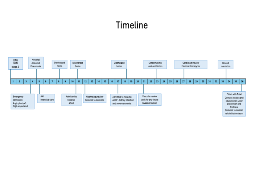
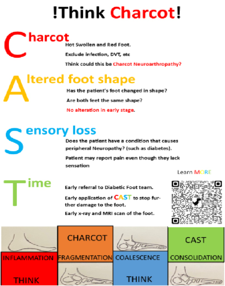
Charcot neuroarthropathy is a significant complication in diabetic neuropathic feet. Without early detection, it swiftly advances, causing considerable deformities in foot architecture and, in severe instances, potentially leading to lower limb amputation (Wukich et al, 2023). However, there is variability in awareness among health professionals regarding the gravity of this condition and its rapid progression (Schmidt et al, 2017).
Here, we present two cases illustrating delayed diagnoses of Charcot neuropathy and the ensuing complications. These cases highlight the consequences of missed or delayed recognition and emphasise the importance of early clinical suspicion and prompt intervention.
In presenting these cases, we aim to highlight the clinical consequences of delayed diagnosis of Charcot neuroarthropathy and emphasise the importance of early recognition, prompt offloading and timely referral to specialist foot services.
Case 1
A 58-year-old man with a history of type 2 diabetes, schizophrenia and prior deep venous thrombosis (DVT) presented to the emergency department with a painful right foot and difficulty weight-bearing. In view of his medical history, DVT was initially suspected; however, Doppler ultrasonography excluded thrombosis.
Plain radiography of the foot demonstrated irregularity of the cuneiform bones, although the views were only partially visualised. The reporting radiologist recommended repeat imaging and orthopaedic referral for further assessment.
The patient was reviewed 1 month later by the orthopaedic registrar who made a provisional diagnosis of arthritis of the cuneiform bone and planned to follow the patient up in arrangement in 3 months’ time.
The patient presented to the acute medical unit with the same complaint 2 months later. A repeat radiograph was arranged, but he was discharged with pain medication, pending orthopaedic appointment the next month.
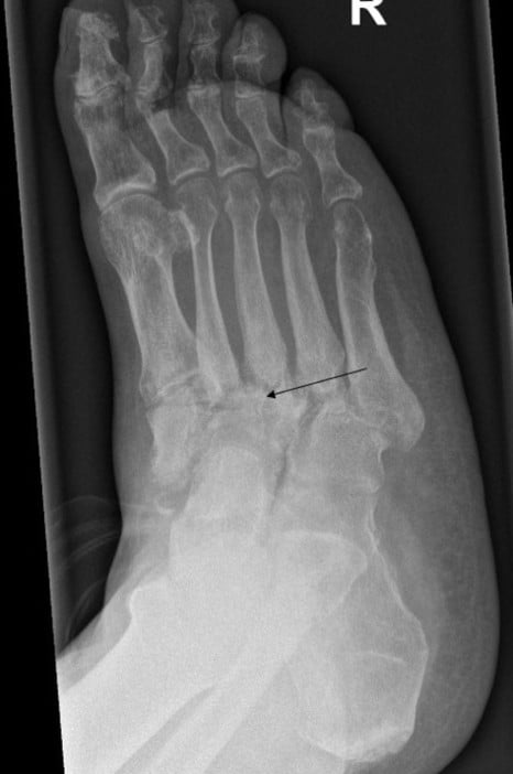
Retrospective review of the second radiograph revealed plantar subluxation of the navicular with disruption of the naviculo-cuneiform joint, consistent with early Charcot neuroarthropathy; however, these findings were not recognised at the time (Figure 1). During the 4-month interval from initial presentation, the patient continued to weight-bear, resulting in disease progression to a Lisfranc fracture, with collapse of the midfoot arch and development of a rocker-bottom deformity. Subsequent MRI confirmed Charcot neuroarthropathy.
The patient was subsequently managed in a high-risk foot clinic and underwent prolonged offloading, including total contact casting followed by an air-cast boot, with a total immobilisation period of 13 months until the condition became quiescent.
Despite these measures, he developed a plantar ulcer over the rocker-bottom deformity, which progressively deteriorated despite antibiotic therapy. The ulcer became infected, necessitating multiple courses of IV and oral antibiotics. Owing to persistent non-healing ulceration and recurrent infection, the patient ultimately underwent a below-knee amputation.
Case 2
A 57-year-old man, previously well, with type 2 diabetes presented to the emergency department with pain and swelling of his left foot. DVT was excluded, and he was discharged with simple analgesia. The patient was reviewed by the orthopaedic resident doctor, who did consider Charcot neuroarthropathy as a differential diagnosis and referred the patient to the foot and ankle specialist, but unfortunately failed to offload the foot. In the subsequent review a week later, the orthopaedic registrar felt that it could be closed infection and decided to act after the MRI scan results.
The patient presented again 1 month later with persistent symptoms and was reviewed by the orthopaedic team. MRI was requested; however, due to scheduling delays, this was not performed until 6 weeks later, at which point it confirmed Charcot arthropathy.
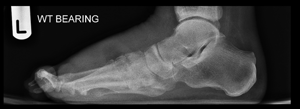
Plain radiography obtained at the initial presentation covered the ankle only; however, when this was repeated later, it clearly demonstrated subluxation of the talonavicular joint with a vertical talus, resulting in a rocker-bottom deformity and associated pes planus (Figure 2).
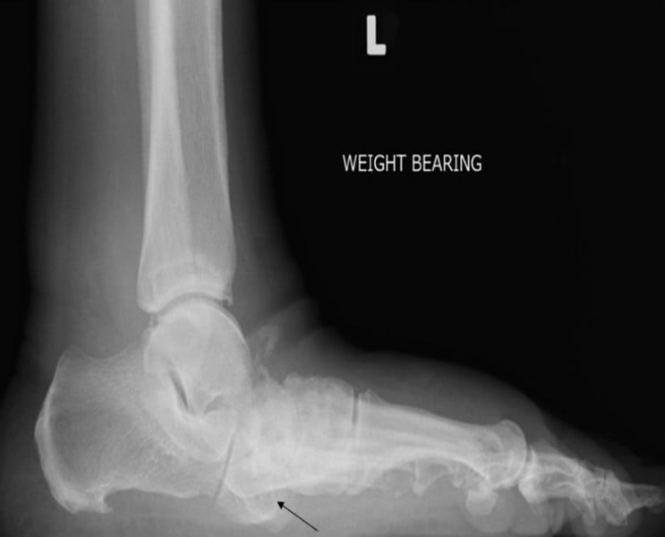
During the 3-month period since the initial presentation, the patient continued to weight-bear, leading to disease progression with structural collapse and complete loss of the medial longitudinal arch.
He was subsequently managed in a high-risk foot clinic, where he underwent prolonged offloading for a total of seven months. His clinical course was complicated by the development of a plantar ulcer requiring regular dressings. The ulcer eventually healed, and he was fitted with custom therapeutic footwear for long-term use prior to discharge to community podiatry services. However, he was later re-referred with recurrent ulceration necessitating further management.
Discussion
Overview
Charcot arthropathy – or neuroarthropathy – was named after the French neurologist Jean-Martin Charcot. The condition was initially described in patients with tabes dorsalis (Sanders et al, 2013). Although, the condition can be seen in other neurological conditions, diabetes is the most common cause worldwide (Dardari, 2020).
This uncommon, but devastating, complication is often overlooked by healthcare professionals (Schmidt et al, 2017). It is not uncommon for clinicians with a special interest in diabetes to incidentally detect inactive Charcot arthropathy in their clinical setting when patients were seen for an unrelated condition (Griffith et al, 1995; Jeffcoate et al, 2000).
Given the degree of neuropathy, patients themselves often may not recollect the events that had led to the change in the shape of their foot. Consequently, their healthcare records may not reflect this either. Hence, the true incidence is not clearly known, but it is estimated that prevalence ranges between 0.3% and 0.53% (Wukich et al, 2023). This increases to 13% in the high-risk population (Suder and Wukich, 2012).
Pathophysiology
The understanding of the pathogenesis of active Charcot neuroarthropathy has shifted from neurotraumatic and neurovascular theories to an imbalance between osteoclasts and osteoblasts, resulting in bone demineralisation, and osteopenia (Rastogi et al, 2021).
The recombinant human parathyroid hormone teriparatide showed significant increase in bone remodelling in chronic CN, but its effects on acute cases are not known (Rastogi et al, 2021). Long-term studies are needed to substantiate its use in clinical practice.
Strict immobilisation and offloading with total contact cast remains the gold standard treatment until date (Wukich et al, 2023). Unfortunately, any deformity that occurs when the disease is active becomes permanent (Jeffcoate, 2015).
What these cases demonstrate
These two cases illustrate the consequences of delayed recognition of Charcot neuroarthropathy, particularly in the acute setting (Table 1). In both instances, patients presented with a hot, swollen foot in the context of diabetes, yet the diagnosis was either not considered or not acted upon despite radiographic abnormalities. Continued weight-bearing during this period resulted in progressive structural collapse, leading to significant deformity.
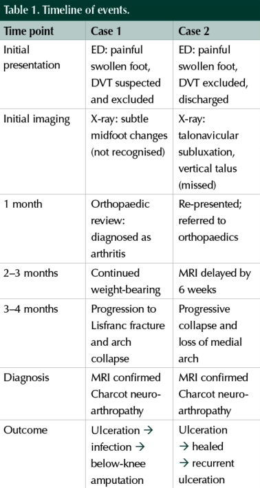
Case 1 highlights the most severe end of the spectrum, culminating in amputation following ulceration and infection. Case 2 demonstrates that even when suspicion is raised, delays in imaging and failure to initiate early offloading can still result in preventable deformity and complications.
Diagnostic challenges and missed opportunities
Missed or delayed diagnosis is a common theme with CN and is seen in as many as 95% of cases before referral to the foot specialist (Schmidt et al, 2017). The average delay is approximately 29 weeks (Pakarinen et al, 2002). In our second case, although the suspicion of CN was raised and MRI scan was booked accordingly, failure to offload promptly resulted in complication.
Chantelau (2005) demonstrated in his small cohort that almost all patients who were misdiagnosed progressed to developing fractures. Left untreated, Charcot neuroarthropathy may progress to fractures, dislocations, deformity, ulceration, infection and even amputation, leading to serious and enduring impacts on quality of life. The diagnostic delay often occurs because Charcot arthropathy mimics common medical conditions such as DVT, cellulitis, gout, osteomyelitis, tubercular rheumatism and complex regional pain syndrome (Rastogi et al, 2022). The average time interval from onset of symptoms to application of total contact cast was 1 month for those who were diagnosed on time as opposed to 3 months in those who were misdiagnosed (Chantelau, 2005).
Importance of early offloading
The most critical learning point from these cases is the impact of continued weight-bearing. Both patients remained fully weight-bearing during the diagnostic delay, which directly contributed to progression of joint destruction and deformity.
Current best practice supports immediate immobilisation and offloading based on clinical suspicion alone, without waiting for confirmatory imaging. Immediate immobilisation and offloading are mandatory when a person with diabetes presents with unilateral foot swelling, increased skin temperature, erythema, and peripheral neuropathy indicative of acute Charcot neuroarthropathy. Suboptimally, offloading in both cases commenced only after MRI confirmation, contrary to best practice guidelines (Wukich et al, 2023).
Once deformity develops, it is largely irreversible and significantly increases the risk of ulceration, infection, and amputation, as demonstrated in case 1.
Mortality and morbidity
The mortality rate for CN varies from 15% to 44% in the published literature due to differences in the baseline characteristics (Chaudhary et al, 2019). A calculated pooled mean 5-year mortality of 29% for CN in 2020, which is comparable to cancer (Armstrong et al, 2020).
Prolonged immobilisation up to 18–25 weeks has been reported in various centres (Game et al, 2012; Rastogi et al, 2021).
Long-term therapy often has its own perils including muscle wasting, reduced mobility, osteopenia, reduced social functioning and significant decline in quality of life (Raspovic and Wukich, 2014). Interestingly substantial skeletal muscle loss occurs within 5 days of disuse (Wall et al, 2014). The considerable impact on everyday function, independence and mental wellbeing experienced by our patients is illustrated in Box 1.

Conclusion
Delayed recognition of Charcot neuroarthropathy can result in rapid and irreversible foot destruction. Any patient with diabetes presenting with a hot, swollen foot should be managed as suspected Charcot neuroarthropathy until proven otherwise. These cases emphasise that early clinical suspicion, immediate offloading, and timely referral to specialist services are critical to preventing avoidable deformity and limb loss.
Consent
Written informed consent was obtained from both patients for publication of clinical details and accompanying images.