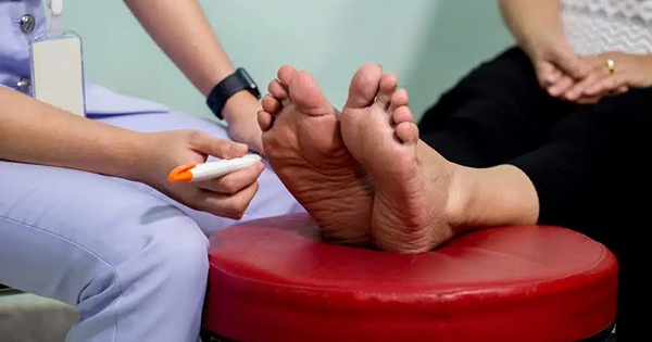
Approximately 15% of patients with diabetes will develop an ulceration and 15–20% will have an ulcer that ultimately leads to an amputation (Frykberg, 1999). The foundation of wound management is wound debridement, removal of non-viable tissue, as failure to do so can result with inflammation, infection and delayed healing. There have been several attempts to determine which if any wound debridement method is superior as it relates to outcomes in wound healing. In 2016, Elraiyah et al performed a systematic review of wound debridement in chronic diabetic foot ulcers (DFUs), determining that the literature supports the utilisation of surgical, autolytic and larval debridement techniques. However, there was low-quality comparative evidence to support one method was superior to another. The authors believe this is because wounds are dynamic and unique, thereby, requiring wound care providers to constantly change their wound care techniques to meet the demands of the ever-changing wound environment.
In this review, the authors aim to take a different approach to determine if the literature provides practical clinical pearls that can aid clinicians in the selection of a particular wound debridement modality and how to employ techniques in a manner that will improve outcomes for patients.
Methodology
The authors sought out to identify the literature reviewing the different wound debridement modalities and techniques. PubMed was searched using an advanced filter to limit the results to clinical trials written in English. The key words included in this search included: “diabetic foot”, “diabetic feet”, “ulcer(s)”, “debridement”, “autolytic”, “biologic(s)”, “collagenase”, “dressing”, “enzymatic”, “lavage”, “maggot(s)”, “mechanical”, “ointment”, “santyl”, “surgery”, “surgeries”, “surgical”, “surgically”, “topically”, “ultrasound” and “versajet”. Inclusion criteria that allowed studies to be evaluated for the final review process included studies that focused on diabetic foot ulcerations with evaluation of a specific wound debridement type as the focus of the study. Studies that were describing non-debridement wound techniques or did not discuss enough relevant wound healing information were excluded.
Results
The PubMed database search yielded 937 studies, including clinical trials, as well as other studies. When clinical trials only were included, the results were narrowed down to 480 studies. After abstract review for potential inclusion in the study, 33 studies were reviewed in detail. Once the studies were reviewed for inclusion/exclusion to the final study, 16 studies were included for final review in this paper. The flowchart describing the study selection process can be seen in Figure 1 and the studies included in our full text review can be seen in Table 1. The studies included provide some insight into demographic information, pertinent diabetes glycaemic control information, debridement type, wound healing rates, ulcer surface area, offloading information and potential blinding and cost data. There were some studies that did not include information in every category, but the majority of the information was discussed in the selected studies.
Discussion
Autolytic wound debridement
The simplest form of wound debridement is allowing the body to create a normal response. This is through natural proteolytic enzymes that degrade necrotic tissue. If using autolytic debridement as the main treatment, the wound should be in an acute state with a healthy granular base (Atkin, 2014). Different topical therapies work with this secondary goal, while strictly providing moisture to the wound bed (Galperin et al, 2015). Many modalities exist, while some overlap with other properties or debridement avenues. A non-exhaustive list would include occlusive dressings, normal saline wet-to-dry or moist-to-dry, hydrogel medical grade honey, topical antibacterial therapies, and different compound formulas. Cost may be consideration with this therapy short term, however, duration of care may offset this when a wound transitions from an acute to chronic state. Studies suggest that adding an enzymatic topical, Clostridial collagenase ointment (CCO; Collagenase Santyl™ Ointment, Smith & Nephew) is actually more cost effective (Tallis et al, 2013; Motley et al, 2018).
Enzymatic wound debridement
Clostridial collagenase ointment is the only Food and Drug Administration (FDA) approved enzymatic agent for wounds and burns (Lantis and Gordon, 2017). This uses the bacteria Clostridium histolyticum to help digest native collagens and remove non-viable debris (Shi and Carson, 2009). This topical form should be sought out for when excessive production of non-viable tissue is overcoming the wound, such as when the wound reaches a chronic state. Collagenase ointment combined with sharp debridement can facilitate the progression of wound closure (Tallis et al, 2013; Motley et al, 2014; 2015; Lantis and Gordon, 2017), especially in recalcitrant wounds (Lantis and Gordon, 2017).
It also has better results when compared to hydrogel combined with sharp debridement (Jimenez et al, 2017). Thus, it could be used to help facilitate sharp debridement and stimulate closure in non-healing wounds. Collagenase could also help bridge debridement appointments and offer pain relief for the sensate patient. Literature suggests collagenase debriders have the ability to reduce local inflammation (Galperin et al, 2015). Though multiple theories exist on the mechanism of action, the in vitro study displayed a decreased in pro-inflammatory mediators (Galperin et al, 2015). This could be a special consideration in diabetic wounds since these wounds experience an exaggerated and prolonged inflammatory reaction (Acosta et al, 2008). Additionally, continual debridement may help prevent wound infections, however, this should be further tested (Motley et al, 2018).
Surgical wound debridement
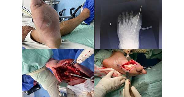
Surgical or sharp debridement has traditionally been considered the gold standard of wound debridement (Acosta et al, 2008). This technique utilises a surgical instrument, such as a scalpel, curette, or tissue nipper to remove devitalised tissue. Its advantage is that it is selective, removing mainly non-vital tissue. Optimal use of surgical debridement requires a very skilled clinician to avoid damage to healthy surrounding tissue. Though quick, effective and commonly recommended, there is insufficient data from randomised controlled trials concerning diabetic foot wounds that prove surgical debridement is superior to any other form of debridement (Edwards and Stapley, 2010). In this literature review, sharp debridement is often combined with other wound debridement types, such as enzymatic and mechanical debridement to improve wound healing time than surgical debridement alone.
Mechanical wound debridement
Mechanical debridement technique physically removes tissue from the wound bed. A pitfall of this modality is that it is non-selective in nature, meaning non-vital tissue, as well as healthy tissue is removed in the debriding process. The most common form of mechanical debridement in regards to DFUs is wet to dry gauze dressings. However, this method can be very time consuming depending on frequency of dressing changes (Kavitha et al, 2014).
More advanced types of mechanical debridement include pulse lavage, hydrotherapy and low frequency ultrasound (LFU) debridement. In a 6-month study, Amini et al (2013) showed LFU in combination with sharp debridement can initially, in the second month of therapy, accelerate wound healing when compared to sharp debridement alone (78% ± 28.7 versus 55.7% ± 31.4, P=0.01) (Amini et al, 2013). However, after 6 months, there was no significant difference between the two study groups. In a pilot study, Yao et al (2014) compared LFU combined with surgical debridement to surgical debridement alone and found that patients that surgical debridement with non-contact LFU as an adjuvant three times a week yielded the best wound area reduction. Larger, well-designed studies are needed to formulate specific conclusions regarding mechanical debridement as it relates to other debridement techniques. Nonetheless, the literature suggests that mechanical debridement combined with surgical debridement may be advantageous for wound healing and wound size reduction.
Biologic debridement therapy
Biologic wound debridement has been a long standing wound debridement modality all over the world with clinical evidence to support its use since the 19th century. One example of this is with use of medicinal maggots, which secrete digestive enzymes that debride by dissolving necrotic tissue, disinfect and thereby promote wound healing. Sherman utilised maggot debridement therapy (MDT) in DFUs unresponsive to conventional therapy/wet to dry (Simmons, 1935; Pavillard and Wright, 1957; Vistnes et al, 1981; Pechter and Sherman, 1983; Mumcuoglu, 2001; Sherman, 2003). Sherman’s study noted that patients that received MDT had 50% reduction in surface area in 9 days versus conventional therapy taking 29 days to reach a 50% surface area reduction. At 4 weeks, MDT had 0% necrotic tissue in wound base versus conventional therapy with 33% necrotic tissue still noted in the base of the wound at 5 weeks. Paul et al (2009) compared MDT versus conventional therapy using Lucilia cuprina instead of the classic blowfly Lucilia sericata in infected DFUs. Lucilia cuprina, which is a more common tropical blowfly, was found to be as effective in removal of necrotic tissue in wounds and stimulating of granular tissue. The end point for this study was a healed DFU or suitable for split-thickness skin graft. The clinical pearl illustrated by both of these studies is that for patients unresponsive to convention therapy or with infected DFUs, MDT should not be considered as a last resort, but sooner in debridement selection process. The demonstration of faster granular tissue promotion is useful for other adjunctive therapies, such as skin graft applications and in overall healing rates.
Other considerations
Wound offloading
Offloading is an integral part of diabetic ulcer healing strategies. Of the studies reviewed, 12 had offloading as a part of the protocol. The type of offloading system use was highly variable ranging from a non-specific offloading device to total contact casting. Based on the inconsistency of offloading systems, the authors could not conclude there was a debridement paired with offloading enhancing technique. They suggest that all DFU care providers use standard of care as it relates to offloading.
Cost
There is increased pressure on cost containment in health care. Management of diabetes in the US is estimated at US$217bn in direct cost in 2017 (Riddle and Herman, 2018) and, as the number of persons with diabetes continues to increase, the authors can expect this cost to continue to rise. The authors noted cost comparison within studies in this review and, of the 16 of studies that met this review criterion, the cost of the debridement modality was discussed in only three clinical trials (Jensen et al, 1998; Tallis et al, 2013; Motley et al, 2014). Future studies on wound debridement should consider cost as this is unfortunately often the basis for selection as providers in the US are relegated to use what insurance companies will cover and patients have the increased burden of making medical decisions based on what they can afford.
Conclusion
The authors reviewed the wound debridement literature to determine if there was evidence to aid practitioners in the selection of one debridement modality over another and useful techniques in implementation of such modality to improve clinical outcomes. The authors recognise the limitations of this review and the evidence in this area. There are few high-quality studies, low patient enrollments, inconsistent head-to-head comparisons, variable randomisation and a lack of consistency in endpoints. In the opinion of the authors, future studies should target designs that eliminate these disparities. However, the evidence does support the general concept that using a wound debridement modality in DFUs is pivotal to decreasing wound healing times.
The CPD module for this article can be found here.