Foot ulceration is a serious complication of diabetes with a reported incidence of 1–3.6% (Ramsey et al, 1999; Boulton, 2008) and a prevalence of up to 25% (Singh et al, 2005). Diabetic foot ulceration is known to result in more hospital admissions than any other diabetes-related complication (Lavery et al, 2006). Infection is a common complication of diabetic foot ulcers and frequently a precipitating factor in amputation (Lipsky et al, 2004). Prevention of infection is, therefore, a clinical priority during wound healing.
The usual approach to diabetic foot ulcer management includes wound debridement, infection control and offloading with regular review. One technology used to assist wound closure and reduce bioburden is topical negative pressure (TNP) therapy. TNP therapy provides negative pressure at the wound surface, thereby lowering oxygen tension, stimulating angiogenesis and removing wound exudate (Banwell and Musgrave, 2004).
Identification and quantification of invasive pathogens is key in the effective management of infection, and thus access to microbiological processing is of paramount importance (Lipsky, 2008). Deep tissue samples are preferred, and repeated sampling is important to verify isolates and provide targeted antimicrobial therapy (Pellizzer et al, 2001). To avoid the overuse of antimicrobial agents, it is important to clinically differentiate between soft tissue infection and colonisation (Nelson et al, 2006). Among people with diabetes, making such diagnoses can be difficult due to the dampening of inflammatory responses (Nelson et al, 2006).
In this article, the authors present the case of Mr X, a 48-year-old man with type 2 diabetes who presented with a non-healing neuroischaemic ulcer to his lateral malleolus. The ulcer was colonised but not infected. A silver foam dressing was used in conjunction with TNP therapy in an effort to avoid infection and achieve wound closure.
Background
TNP has been used to facilitate wound drainage since the 1940s (Vikatmaa et al, 2008). The development of TNP techniques for use on complex open wounds, including the management of diabetic foot ulceration, is more recent (Noble-Bell and Forbes, 2008).
TNP in the clinical setting
TNP therapy involves prolonged exposure of the wound to sub-atmospheric pressure via a closed circuit. The computerised TNP unit generates sub-atmospheric (negative) pressure through a dressing to the wound site, and draws away wound fluid. Dressings are cut to size and applied to the wound and covered with an adhesive drape that maintains an airtight seal. A pressure-sensitive pad placed over a hole in the drape allows the TNP unit to monitor and adjust the pressure being delivered. Tubing connects the dressing to a reservoir to collect the wound fluids that are drawn off. Negative pressures for the purpose of wound therapeutics vary; –75 mmHg is common for use with gauze-based dressings, –125 mmHg for foam dressings (KCI Medical, 2007).
A range of benefits have been associated with the use of TNP therapy in complex wounds (Shi et al, 2003; Saxena et al, 2004; Greene at al, 2006; Morykwas et al, 1997). Briefly, these include:
- Optimisation of blood flow through capillary proliferation.
- Increased rate of formulation of granulation tissue.
- Reduction of tissue oedema.
- Removal of inflammatory by-products contained in exudate.
- Reduction of bacterial count at the wound site.
- Increased epithelialisation.
Dressings for use in TNP therapy
Two types of dressing are used at part of TNP therapy: moist gauze and foam dressings. Silver foam dressings have antimicrobial properties and may be used in colonised wounds. In a moist wound environment, oxidation of the metallic silver results in the sustained release of silver ions that create a barrier on the wound surface and limits the presence of a range of pathogens (Cutting et al, 2009).
Literature review
Review of the literature returns a large number of studies on the use of TNP therapy. This includes three single-centre (McCallon et al, 2000; Eginton et al, 2003; Etoz et al, 2004) and one multi-centre (Armstrong et al, 2005) randomised control trials (RCTs) relating to the management of diabetic foot ulcers with this technology.
McCallon et al (2000), in their single-centre RCT, enrolled 10 participants with postoperative diabetic foot wounds and randomly assigned them to receive TNP therapy (n=5) or control (moist gauze dressings; n=5). The primary outcome was time to satisfactory healing, with the TNP therapy group achieving this in a mean of 22.8±17.4 days, compared with 42.8±32.5 days for the control group. Data on patient satisfaction and the number to achieve wound healing were not provided.
In their cross-over RCT, Eginton et al (2003) demonstrated a significant reduction in wound depth (P<0.05) following TNP therapy when compared with management with moist gauze dressings. The findings of Eginton et al (2003) were supported by Etoz et al (2004) in a larger number of wounds than the previous RCT (n=24 vs. n=7). Etoz et al (2004) reported that the mean surface area of ulcers randomised to receive TNP therapy was significantly less than that of those who received treatment with moist gauze dressings (P<0.05).
Of the four studies, Armstrong et al’s (2005) multi-centre RCT, with 162 enrolled participants, is the most robust investigation of TNP therapy in the diabetic foot. The authors reported that more participants healed (P=0.040), that the rate of wound healing was faster (P=0.005) and that the rate of granulation tissue formation was faster (P=0.002) among those randomised to receive TNP therapy (n=77) compared with control (moist gauze dressings; n=85).
In 2006, Andros et al published the “Consensus statement on negative pressure wound therapy (V.A.C. Therapy) for the management of diabetic foot wounds”, a position statement for the management of diabetic foot wounds using TNP. This document summarised the current clinical evidence, offered guidance on best practice for TNP therapy and highlighted areas for further research.
Case study
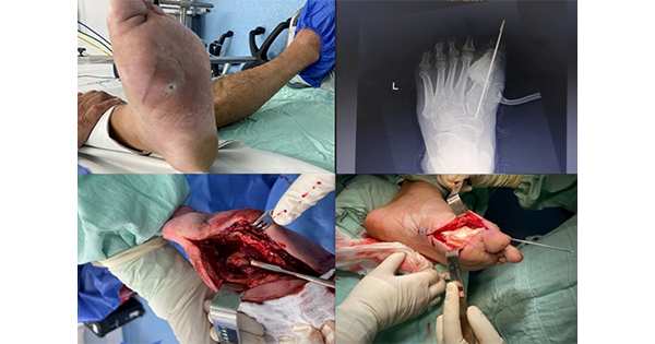
Mr X, a 48-year-old man with type 2 diabetes, presented with a neuroischaemic ulcer over the lateral aspect of the right malleolus (Figure 1). Mr X had undergone orthopaedic surgery to remove a pin in the ankle. The incision site dehisced following removal of sutures, resulting in the ulcer.
Twelve months prior to the current episode of ulceration, Mr X underwent a trans-tibial amputation of his left leg. This was performed due to a non-healing ulcer, as a result of inadequate arterial blood flow to the lower limb.
To preserve Mr X’s remaining intact lower limb, resolution of the current episode of ulceration was a high priority.
Clinical examination
On presentation, Mr X’s ulcer had persisted for 8 weeks. The ulcer had been treated with a range of commercially available dressings, but had shown no indications of healing.
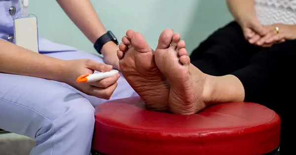
Clinical examination of Mr X revealed peripheral neuropathy of the involved foot, determined by inability to feel a 10-g monofilament or a neuro-tip pen for sharp sensation, and a vibration perception threshold of >25 volts. Peripheral arterial disease was evidenced by the lack of palpable pedal pulses – both posterior and anterior tibial. Mr X’s systolic ankle blood pressure was 100 mmHg. The transcutaneous partial pressure of oxygen at the peri-wound site was <30 mmHg, indicating poor tissue oxygenation.
The ulcer contained a deep central plug of slough at presentation. Following debridement, the ulcer was found to extend to tendon and bone. The ulcer margins were uniformly undermined by approximately 5 mm (note the area of tissue destruction underneath the intact skin of the wound margin in Figure 1). Mr X did not display any of the classic signs of infection (e.g. erythema, warmth, cellulitis, evidence of systemic infection [Lipsky et al, 2004]) and therefore deep tissue samples were not taken.
Treatment regimen
A silver impregnated foam dressing (GranuFoam Silver, KCI Medical, Kidlington) was applied to Mr X’s ulcer in conjunction with TNP therapy at –125 mmHg continuous pressure. A Freedom V.A.C. Unit (KCI Medical) was used to allow Mr X to continue his daily activities applied to the wound. Routine podiatric treatment and dressing change was carried out every 72 hours.
Microbiology
To provide semi-quantitative and quantitative data on the microbiology of the ulcer, wound surface swabs were cultured at weekly intervals. Isolation of aerobes was by inoculation on blood agar, MacConkey agar and meticillin-resistant Staphylococcus aureus (S. aureus) chromogenic agar and incubation for 24 hours at 37°C. For the isolation of anaerobes, specimens were inoculated on neomycin agar and incubated under anaerobic conditions for 48 hours at 37°C. All isolated organisms were identified by conventional microbiological methods. Microbiology reports included an estimation of the number of pathogens according to growth the plate quadrants. Cultures were gram stained for direct examination.
Wound progression
Total bacterial count (the number of bacteria in 1 mL of the sample that can form colonies on blood agar after incubation) was observed to decrease from numbers in the order of 106 colony-forming units/mL to zero culturable colonising organisms by week 6 of therapy (Figure 2).
Enterococcus faecalis (E. faecalis) was the dominant colonist and was isolated from the wound during the first 4 weeks of treatment (Figure 3). Staphylococcus xylosus (S. xylosus) was cultured from wound swabs in weeks 2 and 4 (Figure 4). Both E. faecalis and S. xylosus counts greatly decreased from week 2 to week 5. S. aureus was present only in week 5 (2 × 10³ colony-forming units/mL).
An increase in granulation tissue of approximately 70% was noted by week 6 (Figure 5). Complete healing of Mr X’s ulcer was achieved approximately 6 months after presentation. He remains healed at the time of writing.
Discussion
E. faecalis inhabits the gastrointestinal tract, oral cavity and vagina. The species produces a number of virulence factors and can cause life-threatening infections, especially in hospital settings (Moellering, 1992; Kuriyama et al, 2003). The colonisation of Mr X’s ulcer with E. faecalis, in conjunction with his known vascular insufficiency, was a potentially limb-threatening combination. The eradication of E. faecalis as a colonist during TNP therapy with a silver-impregnated foam dressing was achieved without recourse to antibiotic drug administration.
Conclusion
This case study suggests a role for TNP therapy in conjunction with a silver-impregnated foam dressing in reducing the bacterial load of colonised diabetic foot ulcers to avoid possibly limb-threatening infections.
Acknowledgments
The authors would like to thank Claire Weston, Clinical Marketing Manager, KCI Medical, Kathryn Crawford, Amputee Specialist Nurse, Manchester Royal Infirmary and Stuart Metcalfe, Consultant Podiatric Surgeon, Solihull PCT for their assistance in developing this article.
Conflict of interest
The authors have no conflicts of interest to declare.