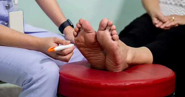
Diabetic peripheral neuropathy (DPN) is the most common diabetic complication and is associated with significant morbidity, including disability, foot ulceration and lower-extremity amputation (Khdour, 2020). DPN is a microvascular complication of diabetes that can affect different nerve pathways, namely sensory, motor and autonomic nerves (McIntosh, 2017). Neuropathy involving the autonomic nervous system, also known as autonomic neuropathy (AN), is a common, but often unrecognised, microvascular complication of diabetes that can present in the form of cardiovascular, gastrointestinal and sudomotor manifestations (Tentolouris and Tentolouris, 2021).
In the lower limb, inadequate sweat gland function, as a result of sudomotor dysfunction in diabetes, is associated with dry skin, itching and anhidrosis, which can contribute to the development of foot problems, including ulceration (Tentolouris and Tentolouris, 2021).
There are several tools available that are used to diagnose sudomotor dysfunction related to AN in the lower limb. However, there is no internationally recommended or recognised standardised tool for this purpose, and it is also unclear if the currently available tools validly measure anhidrosis.
Background
AN in the diabetic foot
The autonomic nervous system is an involuntary system with sympathetic and parasympathetic branches. AN in the diabetic foot is associated with decreased sweating, loss of skin temperature regulation and interruption of sympathetic nerve function (Mulder et al, 2003). Sudomotor dysfunction associated with AN can cause anhidrosis in the feet, making the skin prone to callus, fissures and ulceration (Foss-Freitas et al, 2008; Mascarenhas and Jude, 2014). Autonomic nerve dysfunction, particularly in the lower limb vasculature, also contributes to increased bone resorption from increased local blood flow and arteriovenous shunting (Mascarenhas and Jude, 2014). Arteriovenous shunting can cause weakening of the bones in the foot predisposing to osteopenia, spontaneous fractures and Charcot neuroarthropathy (Vinik and Erbas, 2013; Mascarenhas and Jude, 2014).
In 1995, Young et al undertook seminal research in this area. The authors conducted a cross-sectional study that examined neurological function and bone density in matched groups of neuropathic diabetic patients with and without radiological evidence of Charcot neuroarthropathy. The authors observed reduced bone density in the lower limbs of the Charcot patients compared with the control group (P=0.009), thus identifying that reduced bone strength is likely to increase fracture risk in the feet — an explanation for the initiation of Charcot neuroarthropathy in these patients.
Vinik et al (2003) suggested that disruption of microvascular skin perfusion and sudomotor function may be one of the earliest manifestations of AN in the diabetic foot. Buchmann et al (2019) advised that assessment of the lower limbs in patients with suspected sudomotor dysfunction is of particular importance, because patients may experience early signs of AN, including changes in epidermal moisturisation, anhidrosis and hyperkeratosis of the feet.
Assessment of AN in the foot
Clinical observation of the lower limbs for signs of AN is an important component of a comprehensive patient assessment. However, clinical observation can be subjective and is dependent on the experience and expertise of the clinician. Podiatrists regularly treat patients with diabetes who present with anhidrosis, yet there is no accepted model for identification of anhidrosis or assessing the extent or severity of anhidrosis on the foot (Young et al, 2014).
There are several tests available that are used to detect autonomic nerve dysfunction. Thermoregulatory sweat testing is considered to be the gold standard method for the assessment of peripheral and central sympathetic sudomotor function (Buchmann et al, 2019). Other sudomotor tests include Neuropad® (Trigocare International, Wiehl, Drabenderhöhe, Germany), a diagnostic test device for autonomic dysfunction and early detection of DPN, and Sudoscan (Impeto Medical, Paris, France; Bordier et al, 2016). Both of these detect dysfunction affecting the autonomic fibres.
Sweat glands are controlled by the autonomic nervous system; therefore, an assessment of sweat gland function may serve as a proxy for AN (Malik, 2008). The detection of sweat as a marker of peripheral AN was originally championed by Ryder et al (1988), with the acetylcholine sweatspot test. However, this test was not used widely because it was complex to use and difficult to interpret the results.
Neuropad
Neuropad is a visual indicator test in the form of a plaster containing a blue-coloured complex anhydrous salt – cobalt II chloride. In the presence of moisture, this changes colour from blue to pink, detecting sweating through colour change. It has been proposed as a “simple triage test that can be used to diagnose sudomotor dysfunction and DPN” (Quattrini et al, 2008).
A systematic review and meta-analysis carried out by Tsapas et al (2014) reviewed 18 studies with 3,470 participants and concluded that Neuropad has a sensitivity of 86%. Neuropad is considered an inexpensive, practical, first-line diagnostic screening test for subclinical AN (Spallone et al, 2009; Papanas and Ziegler, 2011).
However, there remain several challenges with its use. Interpretation of findings can be challenging because there is a subjective component to the interpretation of any colour change. Furthermore, the lengthy time needed for patient acclimatisation, setup and application time (15–20 minutes) has resource and cost implications (Tsapas et al, 2014). Neuropad requires moisture detection over 10 minutes, which is longer than nerve conduction testing. The protracted time also leaves the test and findings vulnerable to environmental factors, such as temperature and humidity. Neuropad also has low specificity, with positive and negative likelihood ratios (LR) of LR+=2.44; and LR−=0.22, respectively (Tsapas et al, 2014).
Assessing skin anhidrosis
Anhidrosis refers to the condition in which the body does not respond appropriately to thermal stimuli by sweating (Park and Park, 2019). In the diabetic foot, an absence of sweating can cause changes in epidermal moisturisation, giving rise to anhidrotic skin and hyperkeratosis (Buchmann et al, 2019). Clinicians frequently see presentations of anhidrotic skin in the diabetic foot and give advice regarding the use of emollients to treat it. However, there are currently no validated tools that specifically measure the extent of anhidrosis, or record changes in the presentation of anhidrosis over time for the diabetic foot. Such a tool would allow for a measure of anhidrosis and inform clinicians regarding best practice for treatment of this common condition. A photographic scale to assist in assessment and to improve tissue viability has previously been proposed by Young et al (2014).
Michigan Neuropathy Screening Instrument
DPN is the most significant cause of foot ulceration (Reiber et al, 1999) and is also identified as a major predictor of diabetic foot ulceration (DFU) (O’Loughlin et al, 2010; Alavi et al, 2014). More than 80% of lower-limb amputations occur as a result of DFU or injury, which can result from DPN (Boulton, 2005). The standardisation of diagnostic tools for DPN is essential to support their adoption into clinical practice and inform understanding of the epidemiology of DPN (Mete et al, 2013).
The Michigan Neuropathy Screening Instrument (MNSI) was developed as a screening tool for DPN by the Michigan Centre for Diabetes Translational Research (MCDTR). It is a simple, non-invasive and valid measure of distal symmetrical peripheral neuropathy when compared with gold standard diagnostic testing, including standardised electrophysiology examinations (Feldman et al, 1994; Herman et al, 2012). The MNSI involves two separate assessments: a 15-item self-administered questionnaire and a lower-extremity examination that includes inspection and assessment of vibratory sensation and ankle reflexes.
Herman et al (2012) evaluated the performance of the MNSI in detecting DPN in patients with type 1 diabetes (T1D) and found it had sensitivity of 61% and specificity of 79%. The MNSI is recommended as an accurate validated tool and is a simple and useful screening test for DPN (Lunetta et al, 1998; Moghtaderi et al, 2006).
The use of clinical examination results from MNSI alone has been used as a diagnostic tool for DPN (Lunetta et al, 1998; Moghtaderi et al, 2006; Jaiswal et al, 2013).
Researchers acknowledge the limitations of the MNSI. The scores are known to decrease in the presence of subclinical DPN, causing low sensitivity (Moghtaderi et al, 2006). A further limitation is its inability to screen for autonomic neuropathy (Lunetta et al, 1998; Moghtaderi et al, 2006). Nonetheless, both sets of authors concluded that the MNSI is an accurate validated tool and a simple and useful screening test for DPN (Lunetta et al, 1998; Moghtaderi et al, 2006).
Limitations of current tools
While several tools exist to diagnose sudomotor dysfunction related to AN in the lower limb, there is no internationally recommended or standardised tool for this purpose. A significant limitation of sudomotor tests, including Neuropad and Sudoscan, is a lack of research validating them against standardised clinical examination (Papanas and Ziegler, 2014). It is also unclear if the currently available tools validly measure anhidrosis. Further research is warranted. Therefore, in the present study we investigated associations between MNSI scores, a sudomotor test (Neuropad) and clinically scored anhidrosis.
Aim
The primary aim of this study was to identify the relationship between clinician-observed anhidrosis, Neuropad-detected anhidrosis and Michigan Neuropathy Screening Instrument (MNSI) findings.
Methods
Design
A cross-sectional study was undertaken in a community sample of patients with diabetes. MNSI was administered and Neuropad was applied as per manufacturer instructions. In the absence of a validated tool to measure anhidrosis, skin anhidrosis on the feet was recorded as present or not present, based on whether or not it was observed by the lead researcher (OC). Demographic information was also captured.
Ethics
Ethical approval was granted by Galway University Clinical Research Ethics Committee (ref. CA1323).
Sample
Fifty participants with a confirmed diagnosis of diabetes were purposively recruited from a community podiatry service in the West of Ireland. Participants were aged over 18 years and had T1D or type 2 diabetes (T2D) for a minimum of 5 years.
Patients who had a history of disease consistent with the potential for non-diabetes-related neuropathies, such as malignant disease, alcohol induced neuropathy and vitamin B12 deficiencies, were excluded.
Procedure
MNSI
Permission was sought and granted from MCDTR for the use of the MNSI in this research. The organisation provides instructions for the use of the MNSI on its website and OC adhered to these instructions during the data collection procedure (https://medicine.umich.edu/sites/default/files/downloads/MNSI_howto.pdf).
The MNSI comprises two parts, an examination (MNSIE) and questionnaire (MNSIQ). The MNSIE is undertaken by the clinician and involved observation of skin dryness, callus, infection and deformities, recording of ulceration, testing of ankle reflexes, vibration perception and sensation using a reflex hammer, 128 Hz tuning fork and 10 g monofilament, respectively. The MNSIQ encompasses 15 patient questions which were administered by OC.
Following a review of the literature, MNSIE scores ≥2.5 were considered diagnostic for DPN (Mete et al, 2013). The MNSIQ explores DPN symptoms and a higher score indicates higher DPN risk. A MNSIQ score of ≥4 was used as an indicator of DPN risk (Herman et al, 2012; Jaiswal et al, 2013).
Neuropad
Neuropad manufacturer guidelines were adhered to during data collection. The manufacturer recommends application of Neuropad to the plantar aspect of each foot following removal of footwear and hosiery and a 5-minute acclimatisation at room temperature.
A Neuropad plaster was simultaneously applied to the plantar aspect of each foot between the first and second metatarsophalangeal joint (Malik, 2008). In the presence of moisture, the plaster changes colour from blue to pink (Figure 1).
Colour change and the length of time taken for the colour change was noted, to a maximum of 10 minutes, and recorded. Results were considered normal if there was a complete change from blue to pink and abnormal if there was a partial change or no change at all.
Results
Demographics
Fifty participants were recruited into the study. All participants were Caucasian and of European background. Table 1 shows their demographic characteristics.
Prevalence of DPN
An MNSIE score ≥2.5 indicated DPN (Mete et al, 2013). Results indicated DPN in 66% of participants (n=33).
Prevalence of AN
The prevalence of AN indicated by Neuropad was 72% (n=36) in the total sample.
AN was confirmed in 10% (n=5) of participants where there was no colour change in the Neuropad after 10 minutes. A partial colour change occurred in 62% (n=31) of participants, indicating some degree of anhidrosis and, therefore, AN.
No and partial colour change were combined to report prevalence of AN. Full colour change to pink occurred in 28% (n=14) of participants, indicating no anhidrosis and ruling out AN.
A total of 66% (n=33) of the sample were diagnosed with DPN. In this subgroup, 76% (n=25) were diagnosed with both DPN and AN, the majority of whom (67%, n=22) had a partial colour change with Neuropad (Table 2).
Prevalence of observed anhidrosis
The prevalence of observed anhidrosis in the feet of the total sample was 42% (n=21). Of the 66% (n=33) of the study population confirmed with DPN, 52% (n=17) had observed anhidrosis, whereas in those who were not diagnosed with DPN the majority (76%, n=13) did not have observed anhidrosis either (Table 3).
Relationship between DPN and AN
For the purposes of analysis, the Nueruopad results for no and partial colour change were combined. This was considered reasonable because both these results indicated the presence of some level of AN. In this study, 72% of participants had a Neuropad result indicating AN (Table 2). Chi-square analysis was used to examine the association between the two nominal variables and no statistically significant relationship between DPN and AN was found (χ2=0.89, df=1, P=0.765).
Relationship between DPN and observed anhidrosis
Pearson chi-square analysis was used to examine the association between DPN and observed anhidrosis. The chi-square test was marginally significant (χ2=3.61, df=1, P=0.058), suggesting that the association is approaching significance.
Relationship between AN indicated by Neuropad and observed anhidrosis
The association between observed AN and Neuropad is shown in Table 4. The chi-square test was not significant (χ2=0.807, df=2, P=0.668), suggesting that AN indicated by Neuropad and observed anhidrosis were not associated. A chi-square test using the combined categorical variable for Neuropad (i.e. combining no colour change and partial colour change) was also not significant (χ22=0.739, df=1, P=0.390).
Relationship between DPN diagnosed by MNSIE and MNSIQ
DPN was confirmed in 32% (n=16) by both clinical examination and questionnaire. However, 34% (n=17) had DPN confirmed by clinical examination but not by questionnaire, i.e. clinical signs were present, but participants did not report symptoms. Meanwhile, 4% (n=2) had no clinical signs of DPN, but MNSIQ indicated DPN, suggesting subclinical DPN was present. Finally, 30% (n=15) did not have clinical signs or symptoms of DPN.
Statistical tests to examine the correlation between MNSIE and MNSIQ were calculated using three types of correlation: Pearson correlation: r=0.333, P=0.009; Kendall’s tau: rtau=0.326, P=0.004; Spearman’s rho: rs=0.374, P=.004. All three correlations were significant.
Chi-square analysis examined the association between MNSIE and MNSIQ. The chi-square test was significant (χ2=6.566, df=1, P=0.010), indicating that the MNSIE result is associated with the MNSIQ. The 89% (n=16) of participants identified by MNSIQ as high risk were confirmed as having DPN by MNSIE, suggesting very good sensitivity of MNSIE. Additionally, the 53.1% (n=17) of participants who were not identified as high risk by MNSIQ were confirmed as having DPN by MNSIE, suggesting a high specificity of MNSIQ, i.e. true negative rate.
Discussion
The primary aim of this study was to identify the relationship between clinician-observed anhidrosis, Neuropad-detected anhidrosis and MNSI findings.
Prevalence of DPN
The prevalence of DPN varies widely, ranging from 8% to 70%, according to the population studied and the diagnostic criteria used (Chicharro-Luna et al, 2020). In this study, 66% (n=33) of participants were confirmed to have DPN. Of this subgroup, 76% (n=25) were identified as having AN by Neuropad. This is slightly higher than the entire study group prevalence of 72%, which might have been anticipated given that AN is a key element of DPN and can present subclinically. However, statistical analysis revealed no association between DPN and AN indicated by Neuropad (P=0.765). This result may be explained by the small study size – a larger sample may have provided statistical support to the findings. As the p value is approaching significance, a type II statistical error should be considered, whereby there is a failure to reject a false null hypotheses due to small sample size.
The prevalence of 66% in this study is within reported ranges in the literature, and corresponds highly with the robust research carried out by Dyck et al (1993) in the Rochester Diabetic Neuropathy Study. However, several large-scale studies investigating the prevalence of DPN in people with diabetes report prevalence rates of around 30%; a cross-sectional study by Young et al (1993) studied 6,487 patients with diabetes and found an overall prevalence of DPN of 28.5%. The EURODIAB IDDM Complications Study involved the examination of 3,250 patients with diabetes across 31 centres in 16 European countries. The prevalence of diabetic neuropathy across Europe was reported as 28% without any geographical differences (Tesfaye et al, 1996). A further cross-sectional study involving 8,757 patients recruited from 109 outpatient diabetes clinics in Italy reported a prevalence of 32.3% (Fedele et al, 1997).
The higher prevalence reported within our study is likely to be due to the setting and population under investigation; our study took place within a podiatry clinic and included patients who regularly sought podiatry treatment. Therefore, the population under investigation were more likely to have clinically detectable neuropathy.
Prevalence of AN
In the current study, a prevalence of 72% of AN in the feet of the entire study group was reported, which is higher than the prevalence of 60% recorded in the literature. Using Neuropad, Eckhard et al (2007) reported a prevalence of 60% but only patients with T1D were assessed. The higher prevalence of AN reported in this study relative to existing research may be explained by issues using Neuropad. The manufacturer recommends that Neuropad is used following removal of footwear and hosiery and after 5-minute acclimatisation of the feet at room temperature, although no minimum room temperature is advised. Room temperature was not checked or recorded during the data collection period and temperature in the clinic may have been variable.
Much of the research into Neuropad validity was conducted in Greece, which has a warmer climate than Ireland. It should be noted that data collection for this study took place during the winter months. A systematic review and meta-analysis conducted by Tsapas et al (2014) excluded Greek research, in an attempt to remove climate and temperature as variables, and confirmed diagnostic accuracy remained intact. However, less damp and cold environments may influence the responsiveness of Neuropad because the skin may respond more promptly when tested in a warmer environment.
The literature describes a complete Neuropad colour change as a normal response, with partial or no response as abnormal (Liatis et al, 2007). Time until complete colour change of Neuropad is associated with severity of DPN (Papanas et al, 2005). For the purposes of analysis, the Neuropad results for no and partial colour change were combined, which is likely to have led to the inflated prevalence of AN of 72%. A central weakness in Neuropad is the difficulty in correctly classifying the colour change from blue to pink.
Only 10% (n=5) of participants had no change in colour indicating AN and 90% demonstrated colour change; of these 62% (n=31) recorded a partial change. However no guidelines are provided by the manufacturer to assist the clinician in determining the degree of colour change, making it highly subjective and liable to clinician error and bias.
This difficulty has previously been described by Ponirakis et al (2015), who recommended that the efficacy of Neuropad would be improved if a continuous output scale as opposed to a categorical scale be used. They proposed use of a sudometric app to address this issue. The difficulty in objectively classifying colour changes in the Neuropad results may have distorted overall study findings.
Prevalence of observed anhidrosis
The prevalence of observed anhidrosis was 42%. It would be reasonable to anticipate that if the skin appeared dry on observation then Neuropad would also be expected to indicate anhidrosis. However, results were not conclusive (P=0.668), but as this result is approaching significance, a possible type II statistical error should be considered. Of the 21 participants with observed anhidrosis, only 14% (n=3) had a Neuropad result confirming anhidrosis and 57% (n=12) had a partial result, suggesting some anhidrosis. Interestingly, 29% (n=6) of those with observed anhidrosis showed a conflicting Neuropad result, despite clinical observation of dry skin. Possibly a larger sample size with greater power may yield clearer results.
Similarly, conflicting results are reported in the 58% (n=29) of participants who presented with no observed anhidrosis. Neuropad results ruling out anhidrosis agreed with no observed anhidrosis in only 27% of participants (n=8). However, the majority (66%, n=19) had partial colour change with Neuropad, suggesting some anhidrosis and AN, even though anhidrosis was not observed by the researcher.
No research was found in the literature comparing Neuropad-indicated anhidrosis and observed anhidrosis. Contradictory evidence was reported, where 7% (n=2) of participants presented with no observed anhidrosis, but Neuropad results indicated anhidrosis. This could be interpreted as Neuropad being more sensitive to detecting anhidrosis even when nothing is visible to the eye.
In this study, we found no statistically significant relationship between DPN indicated by MNSI and observed anhidrosis, as recognised by Lunetta et al (1998). This may be due to the inability to screen for autonomic neuropathy with the MNSI. This may also be attributed to the subjectivity of observing skin changes. Tinea pedis is common in people with diabetes and is caused by Trichophyton rubrum which typically presents with a “moccasin” distribution that may mimic anhidrosis (Penzer, 2005). Tinea pedis can be easily confused with anhidrosis related to AN.
Strengths and limitations
This study provides information regarding prevalence of DPN, AN and anhidrosis in the feet of patients with DM, which, to the best of the researchers’ knowledge, was previously unavailable in Ireland. This information is valuable to healthcare professionals in planning resource allocation and in providing a benchmark against which trends can be monitored. The 66% prevalence of DPN in this group is high. It suggests that the group are selected and this is probably because they were already attending podiatry. Prevalence figures may not be generalisable to the wider population of people with diabetes and DPN and, therefore, the authors’ findings should be interpreted with caution.
Data collection was carried out in a community-based podiatry clinic in the West of Ireland that provided access to a suitable sample population. It is reasonable to infer that results from this study can be applied in an Irish context, as the sample is representative of people with diabetes attending podiatry clinics in Ireland.
All data was collected by one researcher, thereby negating any intra-observer variability. However, as discussed, difficulties were experienced classifying Neuropad colour change and cool Irish temperatures may have affected results.
The small sample of 50 participants limits the strength of the statistical tests used and must be considered when generalising findings to the wider population. A larger sample may reveal more statistically significant findings.
Recommendations for future studies
Further research is required to validate sudomotor methodologies, including Neuropad and Sudoscan, against standardised clinical examination.
We recommend the development of a visual colour scale to assist interpretation of Neuropad results. Furthermore, contradictory results between Neuropad confirmed anhidrosis and observed anhidrosis merits further exploration.
There is no accepted model for healthcare professionals to record or measure foot anhidrosis. There is a need for a validated tool to assist podiatrists in the recording and measuring of skin anhidrosis in the feet of people with diabetes.
Conclusion
This study has answered specific research questions relating to the prevalence of DPN, AN and anhidrosis in the feet of the patients with diabetes in the West of Ireland. This previously unavailable information may be useful when planning and allocating resources.
The MNSI tool offers potential for assessment of (early) symptoms of subclinical DPN when clear clinical signs have yet to present in the feet. Despite existing evidence regarding the value of Neuropad, results were not clear in this study. A more standardised procedure for the use of Neuropad, including monitoring room temperature, and development of a visual colour scale to assist interpretation of Neuropad results is warranted. Furthermore, there is a need for a validated tool to measure anhidrosis in the feet of people with diabetes.


