Foot infections are important complications of diabetes (Most and Sinnock, 1983). Osteomyelitis in particular may be a chronic indolent process leading to morphological changes, draining sinuses, and possibly resulting in amputation of a digit or an entire lower extremity. The most frequently involved bones are the metatarsals and phalanges (Bamberger et al, 1987).
Osteomyelitis
Osteomyelitis is defined as a suppurative inflammation of bone sometimes extending to the bone marrow. Several classification systems exist for osteomyelitis (Waldvogel et al, 1970; Cierny and Mader, 1984).
In ‘haematogenous’ osteomyelitis, bacteria are delivered to the site of infection via the bloodstream. In ‘contiguous focus’ osteomyelitis, however, a local infection has spread to involve the underlying bone.
Osteomyelitis can be further subdivided into acute and chronic forms. Acute contiguous focus osteomyelitis is frequently observed in the foot of the person with diabetes. Local trauma as a consequence of peripheral neuropathy leads to a skin or soft tissue injury, which may become acutely or secondarily infected (Pecoraro et al, 1991; Reiber et al, 1992). A neutrophilic vasculitis secondary to the soft tissue infection ensues (Edmonds, 1999). This leads to breakdown of the soft tissue envelope surrounding the bone which serves to initiate or perpetuate the process (Cierny and Mader, 1984). Once this acute local process begins, it may progress and persist to become chronic, as it goes unnoticed (due to lack of local sensation).
The key feature of chronic osteomyelitis is infected non-viable bone contained within a compromised soft tissue envelope (Cierny and Mader, 1984).
Diagnosing osteomyelitis
Evaluating the status of bone in the person with diabetes and a foot infection is complicated because established criteria do not exist. However, clinical and laboratory data may help establish the diagnosis. It is important to select laboratory investigations that provide useful answers and are cost-effective.
Clinical evaluation
It has been proposed that clinically the ‘sausage toe’ deformity (Figure 1), due to local soft tissue infection and inflammation and underlying bony changes, is highly suggestive of underlying osteomyelitis (Rajbhandari et al, 2000).
Individuals with soft tissue infections or skin ulcerations that have been present for several weeks are at high risk of contiguous bone involvement, particularly if these lesions are located over a bony prominence (Lipsky, 1997).
The larger and deeper an ulceration is, the more likely that an underlying osteomyelitis is present (Newman et al, 1991). Studies have shown that exposed bone, either viewed directly or detected by gentle probing at the base of the wound, correlates well with presence of osteomyelitis (Newman et al, 1991; Grayson et al, 1995).
Laboratory investigations
Erythrocyte sedimentation rate and C-reactive protein level are both acute-phase reactants. Despite their relatively low sensitivity and specificity, they are frequently used for the detection and monitoring of ongoing bone infections (Carlsson 1978; Newman et al, 1991; Haas and McAndrew, 1996; Sanzen, 1988).
Imaging techniques for the diagnosis of osteomyelitis have a wide range of sensitivity, specificity and positive predictive values. The plain radiograph will only demonstrate bony abnormalities related to osteomyelitis 10–20 days after the bone infection has occurred and 40–70% of bone has been resorbed (Shults et al, 1989; Schauwocker, 1992). When plain radiographs are used, it is also important to obtain a baseline radiograph and a follow-up study after 10–21 days to determine whether the typical bony abnormalities are present, specifically cortical destruction with periosteal new bone formation (Haas and McAndrew, 1996).
Table 1 shows available imaging techniques for identifying osteomyelitis in the feet of people with diabetes. A 3-phase technetium bone scan (Tc99m) is sensitive for diagnosing osteomyelitis but suffers from poor specificity in diabetic foot infections because of frequent false positives caused by overlying soft tissue hyperaemia (Keenan et al, 1989) or bony remodelling from trauma (as may be seen with the Charcot foot). Nuclear imaging techniques may also be confounded by profound ischaemia of the lower extremity.
Microbiology
Appropriately collected specimens may help guide focused antimicrobial therapy to treat the infection and prevent the emergence of antibiotic-resistant microorganisms (Tentolouris et al, 1999). Foot infections in people with diabetes are frequently polymicrobial (Louie et al, 1976; Sapico et al, 1980; Wheat et al, 1986). Initially though, the most frequently encountered microorganisms are Staphylococcus aureus and Streptococcus pyogenes. When tissue at the ulcer base starts becoming devitalised, Gram-negative bacilli and anaerobes appear and may play a role in the underlying osteomyelitis.
Superficial swabs of ulcers overlying a focus of osteomyelitis are frequently unreliable. Deep swabs from sinus tracts, deep tissue curettage and bone biopsy specimens are more reliable at detecting the microorganisms causing the underlying osteomyelitis.
A combined approach
Several algorithms exist for the diagnosis and management of diabetic foot osteomyelitis (Haas and McAndrew, 1996; Lipsky, 1997). If osteomyelitis is suspected, the most rational approach is to combine clinical evaluation and diagnostic investigations. Figure 2 demonstrates a simplified algorithm utilising this approach. The first stage of the evaluation must be the physical examination, establishing whether a ‘sausage toe’, and/or a draining sinus or ulcer exist. If so, then it is determined with a sterile probe whether bone can be palpated at the base of the lesion. If yes, a diagnosis of presumed osteomyelitis has been established and treatment may be initiated accordingly. If no, but clinical suspicion persists, a plain radiograph should be obtained at baseline and after 10–21 days. In the intervening period, osteomyelitic changes should be visible radiographically. Therapy may be initiated pending the follow-up plain radiograph. If the follow-up radiograph is unhelpful and suspicion of osteomyelitis persists, a 3-phase technetium bone scan combined with a gallium scan or white blood cell study may be warranted (Keenan et al, 1989). These combinations yield improved specificity while maintaining sensitivity.
It has been suggested that extensive non-invasive investigations generate significant expense without significant improvement in health outcomes for people with diabetes in whom foot osteomyelitis is suspected (Eckman et al, 1996). The most appropriate antimicrobial regimens are those guided by the results of appropriately collected specimens.
Adjunctive measures
When assessing the diabetic foot, it must be remembered that the status of the bone is influenced by surrounding environmental factors, including:
- Vascular status of surrounding tissue: the status of circulation must be evaluated, as adequate circulation is necessary for the resolution of infection and promotion of wound healing (European Working Group on Critical Leg Ischemia, 1992; Hill et al, 1999)
- The presence of an overlying ulceration
- Footwear: adequate footwear facilitates healing of local ulcerations by minimising ongoing trauma.
The entire patient must also be considered, specifically: metabolic status, particularly glycaemic control; renal function; economical factors; psychological factors; and tobacco consumption.
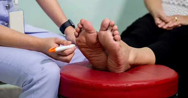
All of these factors will influence the ultimate outcome of the foot ulcer and the underlying bony lesion. In addition to the investigations outlined above, it is critical to establish the presence and extent of neuropathy as the loss of pain perception may lead to increased susceptibility to mechanical and thermal trauma which may adversely affect the local environment.
Antibiotic management
Unlike skin and soft tissue infections which usually resolve with short courses of antimicrobial therapy, management of bone infections requires prolonged courses of treatment. In the introductory article to this journal’s antibiotics series, Lipsky (1999) asked several questions about the optimal management of diabetic foot osteomyelitis. The following discussion is based on these questions.
The best answers would be provided by prospective randomised double blind placebo trials. At the moment, most of the existing data are from retrospective or observational studies.
Duration of treatment?
The optimal duration of antimicrobial therapy is not known. However, the traditional approach has been to provide 4–6 weeks of parenteral therapy (Norden, 1988), or to give an ‘induction course’ of 1–2 weeks parenteral therapy followed by prolonged courses of oral therapy (Bamberger et al, 1987; Norden, 1988; Venkatesan et al, 1997; Pittet et al, 1999). Several existing reports document prolonged courses of oral antimicrobial therapy for the management of diabetic foot osteomyelitis (Wilson and Kauffman, 1985; Peterson et al, 1989). A recent study in our facility revealed that of 128 episodes of osteomyelitis, 80% were successfully treated with oral antimicrobial therapy, remaining relapse-free at one year (Figures 3a and 3b).
Choice of antimicrobials
The choice of antimicrobial therapy should be guided by the microorganisms recovered from bone biopsy specimens, fragments of debrided bone or from deep draining sinus tracts if tissue specimens are not available. The route and type of agent will depend upon the status of the patient and the bone. In clinically stable patients, for instance, a chronic, non-limb threatening infection can probably be managed with a prolonged course of oral antimicrobial therapy. However, an acute limb threatening infection would require parenteral therapy, possibly followed by a prolonged course of oral antimicrobial therapy.
Table 2 shows the antimicrobial agents routinely used in our facility for both parenteral and oral therapy of diabetic foot osteomyelitis. From our experience, the most frequently used antimicrobial regimens are trimethoprim/sulphamethoxazole 960mg po bid combined with metronidazole 500mg po tds or ciprofloxacin 500mg po bd and clindamycin 300mg po qds or, alternatively, amoxycillin-clavulanic acid 500mg po tds. The choice for oral therapy is based on patient tolerability and bioavailability in soft tissue and bone levels.
Although antibiotic choices appear intuitive for the management of diabetic foot osteomyelitis, the process should extend beyond merely ‘matching the bug to the drug’ as not all antimicrobial agents achieve satisfactory bone levels. It is therefore prudent to discuss antimicrobial choices with either an infectious disease or microbiology consultant to ensure that optimal therapy is selected. It is important to note that although multiple pathogens may be recovered from a diabetic foot wound not all are invasive pathogens requiring treatment.
Surgery
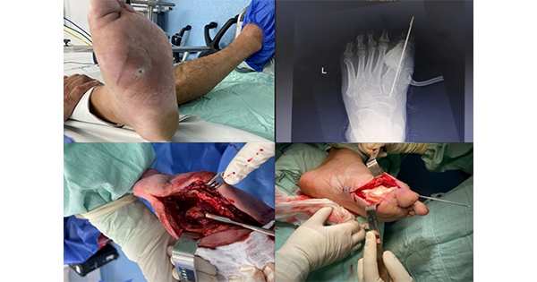
Adequate peripheral circulation is necessary for the healing of skin, soft tissue and bone infections, as well as wounds, in the foot of the person with diabetes. Although antibiotics are highly effective, their efficacy is limited in situations when ischaemic non-viable bone is present or large areas of bone are exposed.
In areas such as in the pulp spaces of hammer toes, where repeated trauma occurs, ongoing soft tissue trauma leads to tissue loss and in many situations osteomyelitis cannot be easily treated.
Several authors have shown that, provided circulation is satisfactory, toe amputation for osteomyelitis in the person with diabetes is cost- and time-effective (Johnson et al, 1987; Benton and Kerstein 1995; Ha Van et al, 1996; Kerstein et al, 1997). Surgery must be undertaken with caution in the neuropathic diabetic foot as amputations will lead to abnormal foot biomechanics and the creation of new abnormal bony pressure points potentially leading to new ulcerations (Edmonds, 1999). The optimal duration of antimicrobial therapy after debridement or amputation of an osteomyelitic digit is not known. Intuitively, if the entire osteomyelitic digit has been resected, only a brief course of antimicrobial therapy is necessary to ensure that local tissue infection does not ensue. If debridement of necrotic bone is undertaken, a more prolonged course of therapy is indicated to prevent invasion of the healing bone edges by bacteria in the surrounding soft tissue.
Conclusions
Once the diagnosis of osteomyelitis has been established in the diabetic foot, it is imperative that available microbiology be used to help guide antimicrobial selections. Diabetic foot osteomyelitis can be successfully treated with prolonged oral antimicrobial therapy. The optimal duration of therapy is not known and can only be established using randomised trials.
Adjunctive measures may be necessary to ensure adequacy of circulation, weight displacement from open ulceration and bony pressure points and debridement of non-viable tissue and bone.
Acknowledgements
The author would like to acknowledge Ms Carolyn Cooper for her valuable secretarial skills in the preparation of this article.


