In order to ascertain risk of chronicity, clinicians should use initial patient assessments to identify wound aetiology and patient comorbidities. However, although clinicians can usually identify problems at initial assessment, wounds often become chronic (defined by a failure to heal within 4 to 6 weeks [HAS, 2016]) if they are not managed effectively for a period of time, for example, due to impeded referrals to a specialist clinician (Oyibo et al, 2001). However, some wounds may from the outset present chronic features, such as diabetic foot ulcers, venous leg ulcers or pressure ulcers. Certain patient groups are more likely to develop a chronic wound due to their multiple comorbidities (i.e. uncontrolled diabetes, peripheral arterial disease, chronic venous insufficiency or recurring foot ulceration).
One such group is patients with diabetes, of whom an estimated 10% will experience a diabetic foot ulcer (DFU) at some point (Kerr, 2012). DFUs are considered challenging due to the presence of diabetes and other comorbidities, and pose a challenge to all practitioners involved in their care; they are often complicated by ischaemia, neuropathy, poor diabetes control and increased risk of infection. Treating DFUs as quickly and appropriately as possible can reduce their longevity (Oyibo et al 2001), whilst positively impacting quality of life.
A DFU has been likened to a ‘foot attack’ – a foot ulcer or infection failing to heal and leading to potential amputation (Diabetes UK, 2013) – and is a major event in the life of a diabetic person, indicating serious disease and the possible presence of comorbidities such as cardiovascular disease (Brownrigg et al, 2012). Every week in England there are over 120 amputations in people with diabetes (Diabetes UK, 2013). Successful treatment to prevent amputation involves a multi-disciplinary, holistic approach, including: providing effective wound care, maintaining optimal diabetes control, restoring adequate circulation, controlling infection and implementing pressure relief. The patient should, of course, be at the centre of this care (NICE, 2015; Wounds International, 2013).
DFUs pose a major burden to the healthcare system, with foot complications accounting for 20% of the total NHS spend on diabetes, equating to approximately £650 million every year (Kerr, 2012). Importantly for the patient, DFUs also have a great impact on quality of life, affecting physical, psychological and social wellbeing. The need for frequent dressing changes and an inability to work or lead a normal life can be life-altering.
Assessment of diabetic foot ulcers
Identifying the aetiology of a DFU is essential to determine appropriate clinical management. They are usually classified as neuropathic (a lack of protective sensation), ischaemic (poor peripheral circulation) or neuro-ischaemic (both reduced sensation and circulation). Loss of protective sensation is associated with a seven-fold increase in risk of ulceration (Singh et al, 2005) and is seen in the majority of patients with a DFU. Patients with this sensory loss are vulnerable to physical, thermal or chemical trauma, which, due to the reduced awareness of pain, can lead to ulceration. An ever-increasing number of DFUs have an ischaemic component, with up to half of wounds complicated by the patient having an inadequate blood supply (Hinchcliffe et al, 2012; Huliberts et al, 2008).
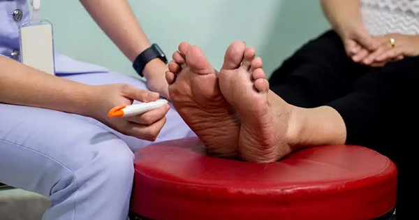
It is crucial to assess the patient’s neurological and vascular status using, at minimum, palpation of foot pulses, application of the 10g monofilament and a visual examination. It is also important to look for signs of infection, which can be difficult with DFUs, where the classic signs of infection (redness, heat and swelling) are often masked due to poor arterial supply and loss of sensation. The International Working Group on the Diabetic Foot and Infectious Disease Society of America’s criteria for recognising and classifying diabetic foot infection are useful, as they clearly describe how to classify severity of an infection. Use of deep wound swabs, soft tissue and bone cultures in clinically infected wounds can assist by identifying the causative pathogen and sensitivities (Lipsky et al, 2012).
Foot deformity and high plantar pressures are also often seen in patients with neuropathy and are common causative factors for DFUs (Singh et al, 2005); clawing of the toes, muscle wasting, high arch, hallux valgus/rigidus and gait changes (e.g. ataxic and Charcot neuroarthropathy) are all seen in patients with neuropathy (Bakker et al, 2012). In patients with peripheral neuropathy, offloading is a crucial piece of the ‘jigsaw’ of wound management, with the total contact cast often cited as the gold standard (Armstrong et al, 2004). These are not suitable for all patients and are contraindicated in the presence of ischaemia and infection, where alternatives like removable casts, Scotchcast boots, Softcasts and walking aids or wheelchairs can be used (NICE, 2015; Armstrong et al, 2004).
Matrix metalloproteinases and their role in wound healing
Matrix metalloproteinases (MMPs) are part of a group of metalloproteinase enzymes that are integral to the healing trajectory (Martin, 1997; Singer and Clark, 1999). The protease groups involved in wound healing are MMPs (MMP-1, MMP-2, MMP-8 and MMP-9) and the serine protease human neutrophil elastase (HNE) (Gibson et al, 2010). Combined, these remove any damaged proteins and assist in cell migration and tissue remodelling, while regulating growth factors.
Inactive MMPs are activated by the presence of proteases and HNE; the resultant MMP activity is then directly influenced by the tissue inhibitors of metalloproteinases (Rohl and Murray, 2013). It is this fine balance between proteases and inhibitors that dictates wound progression, while disruption of this balance stalls the healing process, with elevated protease activity and persistent inflammation trapping the wound in the inflammatory phase and a residual chronic state. Studies examining wound exudate from chronic non-healing wounds confirm this biological irregularity and the destruction associated with high levels of proteases and reduced levels of important growth factors (Troxler et al, 2006). Further studies have attempted to correlate MMP levels within certain populations, wound aetiologies and sample types, with elevated MMPs noted for all chronic wounds, including DFUs. This suggests increased protease levels impede healing in chronic wounds (Gibson et al, 2010).
Poor DFU healing is also associated with changes in the biochemical wound environment compared with non-diabetic wounds; namely, a reduction in growth factors (Flanga 2005), angiogenic response (Maruyama et al, 2007) and migration of keratinocytes and fibroblasts. Additional factors that may influence healing include neuropathy, ischaemia and poor glycaemic control (diabetes-related), and factors such as infection, smoking, nutrition, and non-concordance with offloading devices (non-diabetes-related) (Uccolil et al, 2015).
UrgoStart technology
In all DFUs wounds, it is important to tackle the protease imbalance and therefore allow progression of the healing cascade to occur. Any dressing with the ability to absorb could be classed as having a protease-modulating impact; however, true protease inhibition requires a protease inhibitory compound, such as Nano Oligo-Saccharide Factor (NOSF) (Schmutz et al, 2008). The literature confirms that use of wound dressings that directly influence protease activity in the wound bed leads to better healing outcomes (Lazaro et al, 2016). Protease-inhibiting dressings used in conjunction with optimal care (e.g. diabetes control, skin integrity management and wound bed preparation) should be an early consideration for DFUs due to poor healing rates and associated chronicity. Dressings within the UrgoStart range have been developed for wounds with or at risk of an imbalanced protease equilibrium. They contain the innovative protease inhibitory compound TLC-NOSF (Technology Lipido Colloid), which restores the wound healing imbalance and therefore allows progression of the healing trajectory. The TLC–NOSF dressing range promotes wound healing by inhibiting MMP activity (Schmutz et al, 2008) and in vitro it has been shown to promote proliferation of fibroblasts (McGrath et al, 2014). The TLC combined with NOSF when in contact with wound exudate forms a gel and creates a moist environnment, enabling the key cells involved in the repair process (fibroblasts, keratinocytes) to exert their action.
UrgoStart Contact dressing is a flexible contact layer comprised of a conformable polyester mesh impregnated with hydrocolloid, petroleum jelly and NOSF particles. If there is depth to the wound, UrgoStart Contact should be used to ensure contact of the TLC-NOSF matrix with the wound bed, whereas a superficial wound would benefit from UrgoStart Border, which has additional provision of protection and absorbency, but is not suitable for wounds that require packing.
In 2012, Meaume et al documented results of a double-blind randomised controlled trial for UrgoStart (Meaume et al, 2012). The study assessed the efficacy of the TLC–NOSF within adult patients with non-infected venous leg ulcers. The wounds were assessed fortnightly for 8 weeks, with a primary objective to monitor the Wound Area Reduction (WAR) as a percentage. Patients (n=187) were randomly allocated into two groups, with the differentiator being the presence of the NOSF compound in the UrgoStart group, compared with the control group. A median WAR of 58.3% was seen for UrgoStart group versus 31.6% in the control group, equalling a difference of -26.7% (CI=95%; p=0.002). Clinical outcomes for patients treated with UrgoStart were noted as superior. These results are supported by the case study series presented below.
Case study series
Eleven in-market clinical evaluations were carried out on patients with DFUs by advanced podiatrists with previous experience of using UrgoStart. The objective was to advance chronic wounds that had seen limited progression over previous months to full healing and to prevent longevity of new wounds for patients with multiple comorbidities. At least 27% (n=3) of the patients had been advised that amputation was the next step if healing was not achieved.
Clinical photographs were taken and fully informed patient consent was gained before data inclusion and definitive recruitment. All recruited participants were being managed within the community and supported by regular outpatient visits to the podiatrist or foot services. A bespoke evaluation data collection tool was used to capture detailed baseline information and various evaluation parameters (Table 1). Participants not only included clinicians but also carers and relatives of the recruited patients, so this tool’s design ensured a consistent approach to data collection.
To ensure UrgoStart was used appropriately, 27% of patients (n=3) required a reduction in slough (>30%) prior to commencement of treatment. Once the wounds reached a granulating non-infected state the care was amended. This cohort was representative of the overall DFU population:
- predominantly male
- many with type 2 diabetes
- age range of 55 to 81 years
- mean age: 66 years/median age of 63 years.
An overview of results for the eleven in-market evaluations is provided in Figure 1 and five example case studies, chosen at random from the case study series (Figure 2), are presented below.
Example case studies
Case 1
A 63-year-old male with type 2 diabetes, bilateral Charcot foot, and a number of other comorbidities had been off work, undergone various hospital stays and was frustrated with his regular dressing changes. A previous right-foot partial amputation had healed well. Onset of bilateral DFUs occurred in September 2011.
From September 2011 to February 2013, a variety of dressing regimes were used, including alginates, iodine and silver, but the wound remained static throughout 2012. Offloading devices had also been used, including a total contact inlay and a non-removable cast, but the patient was not compliant with their use and became very unsteady with the cast, so it was discontinued. In February 2013, the patient was admitted to hospital with worsening of his left foot ulceration under the Charcot joint. Surgical debridement was required; on discharge, the dressing regime was a Hydrofiber and a bordered foam 3 to 4 times a week. The patient refused offloading or casting.
UrgoClean was commenced to deslough the wound, which measured 10cm x 20cm, with 80% slough. Thrice weekly dressing changes were planned. After 1 week, the large wound had become three smaller wounds measuring (1) 10cm × 4cm, (2) 4cm × 1.5cm, (3) 1.3cm × 0.8cm. (Figure 3a) Dressing changes were reduced to twice weekly.
One month after commencing UrgoClean, two of the wounds had reduced further in size ([1] 8.4cm × 4.5cm, [2] 1.2cm × 0.3cm) and the third had healed. In May 2015, UrgoStart was commenced with weekly dressing changes; at this point, two of the wounds had healed and the third had further reduced in size: (1) 8cm × 2.2cm (Figure 3b). By late July, the wound measured: (1) 3cm × 0.3cm, representing a 95% wound area reduction in 10 weeks. The patient was extremely pleased when wound healing was achieved (October) (Figure 3c).
Case 2
A type 2 diabetic, insulin-dependent, 55-year-old male was referred for debridement of an extensive surgical wound. At presentation, the plantar surgical wound was almost healed, but was accompanied by a non-healing forefoot ulceration. The wounds were being managed with a retention bandage, a calcium alginate and an absorbent pad, and an offloading slipper cast. The periwound area was macerated and there was a moderate level of exudate. Despite being practically wheelchair-bound since surgery, the patient wanted to walk down the aisle for his wedding and prevent amputation.
This DFU had been present for more than 6 months when UrgoStart Border was commenced and offloading was continued as previous. On initiation of UrgoStart Border (Figure 4a), the wound measured 4.3cm × 3.3cm × 0.1cm. Due to the vulnerability of the foot and previous loss of digits, the specialist podiatrist requested daily dressings for the first 10 days. At day 10, the wound measured 2.1cm x 1.4cm x 0.5mm, equating to a 79% wound surface area reduction (Figure 4b). Dressing frequency was decreased to thrice weekly until full healing occurred at day 56 (Figure 4c).
All clinicians reported ‘excellent’ for all parameters (Table 1). The patient’s partner managed the dressing changes whilst on holiday and reported ease of application and excellent ability of UrgoStart to manage exudate and remain in situ. Throughout this process, the slipper cast was modified in line with offloading requirements and, although the wound remains healed, offloading continues as a preventative measure. The patient achieved his goal of wearing his own shoes to walk down the aisle at his wedding.
Case 3
A 55-year-old, male, insulin-dependent diabetic was under the care of the podiatry team for a previous forefoot amputation that led to numerous offloading requirements. When a new wound developed as a result of his splint prosthesis rubbing, UrgoStart Border was used as the first intention dressing, due to the patient’s history of chronic foot ulcers.
UrgoStart was used in conjunction with offloading to ensure the wound did not become chronic. For this gentleman, remaining at work, which required a large amount of standing, was imperative. Post-new-ulcer formation, the patient used an innovative slipper cast device while his custom-made prosthesis was being modified, and then continued to use this as an in-house slipper when not at work. The modified splint prosthesis was worn at all other times to ensure the patient’s goals of remaining ambulant and at work were achievable without further wound deterioration.
At initiation, the wound measured 2.2cm × 2.5cm × 1mm (Figure 5a), which had reduced by 53% to 2cm × 1.3cm by day 21 (Figure 5b). It is comparatively challenging for patients who wear a prosthesis to achieve full wound healing (Figure 5c); however, in this case, a balance of prosthesis-wearing, the patient remaining at work, and quick wound healing was achieved by way of accurate assessment, appropriate dressing choice and offloading.
Case 4
A 63-year-old type 2 diabetic male with multiple cormorbidities required a right forefoot amputation due to gas gangrene. This was performed on 18.06.15 with discharge on 03.07.15. On referral, the wound measured 12.5cm × 9.5cm and was being managed with absorbent pads, a conformable hydrogel and pressure dressings. The dressing regime was changed to an open-mesh silicone dressing with absorbent pads after deterioration and visibility of exposed tendon and bone. For the following 3 months, a varied range of dressing regimes were undertaken, but the wound had only reduced by 22% during a 12-week period. Throughout the wound healing process, offloading for this patient continued with no adjustments.
When UrgoStart Border was commenced (Figure 6a), the wound measured 12.5cm × 7.4cm, there was 100% granulation and exudate levels were heavy. Four weeks after initiation of UrgoStart Border, the wound surface area had reduced by 44% to 9.4cm × 6.5cm (Figure 6b). The surrounding skin had remained healthy at all times, with only very slight maceration noted at three dressing changes. Although exudate levels remained heavy until month 3, it was managed well by UrgoStart Border. By January 2016, the wound measured 6.7cm × 4.3cm, a 53% reduction in 10 weeks.
By late March, the wound measured 3.3cm × 3.2cm, with 100% granulation and moderate levels of exudate, and was on the appropriate trajectory for full healing, with weekly dressing changes (Figure 6c). Although the forefoot could be classed as a difficult-to-dress area, UrgoStart Border was easy to apply and remained in situ until the next dressing change.
Case 5
A 65-year-old female with neuropathy, who was a heavy smoker and a Hepatitis B carrier but had no DFU history, presented to the podiatry team in January 2016. Her wound initially presented as a cyst, but developed into an ulcer that was being managed by a private podiatrist. When weekly redressing appointments were not available, the patient was referred to the NHS specialist podiatry team. Type 2 diabetes was diagnosed via a blood test in March 2016; this is diet-controlled.
After 14 weeks, the wound measured 0.8cm × 0.5cm × 0.2cm with 100% slough. It had previously been managed with a silver Hydrofiber dressing, foam, tape and offloading padding. This patient was keen to improve her wound as she had been struggling at work, which involved being in a freezer for 8+ hours a day, and her feet were always cold. Her hours had been reduced as she was unable to fulfil her usual role due to the limitations of offloading.
The UrgoStart dressing was used in conjunction with offloading, which remained the same during the wound management process. The podiatrist also issued the patient with some plastazote insoles to provide insulation within her work boots. UrgoStart Border was initiated to ensure the wound did not remain inappropriately in an uncontrolled inflammatory phase. In addition, 10 mm semi-compressed felt was applied to the toe to help offload the pressure.
The wound measured 0.7cm × 0.5cm × 0.1cm after 1 week of using UrgoStart Border and, 4 weeks after initiation (Figure 7a), the wound surface area had reduced by 70% to 0.4cm × 0.3cm × 0.2cm (Figure 7b). The wound continues to improve (Figure 7c), and the patient’s ongoing aim is to return to normal working hours and her full role as quickly as possible.
Conclusion
This paper presents a case study series using UrgoStart products on patients with DFUs, with detailed results of five chosen at random. This TLC-NOSF dressing promotes wound healing by inhibiting MMP activity and promoting proliferation of fibroblasts. The authors recognise the limitations of a small-scale clinical in-market evaluation, but believe the findings are relevant for publication. It is reported that UrgoStart is easy to use, and patients and relatives can apply the dressings themselves. Within a resource-limited NHS, using a safe and effective dressing that is easy to apply has clinical and economic advantages. Moreover, while the focus of this paper is on patients with DFUs, the results are transferrable across wound care.


