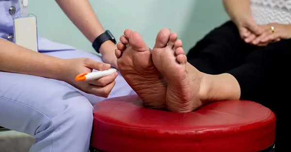
Measurement of vibration perception threshold (VPT) in a clinical environment is an essential element of any neurological examination and provides a routine method of screening for peripheral neuropathy (Young et al, 1993; Merriman and Tollafield, 1995). Diabetic neuropathy affects approximately 30% of the diabetes population (Kumar et al, 1994) and sensation loss may be a very sensitive indicator (Coppini et al, 1998). Measurement of VPT, which reflects large nerve function, is a form of quantitative sensory testing that is strongly associated with various neuropathic indices (Franklin et al, 1990; Kahn, 1992). Consequently it is one of the main diagnostic measurements used in clinical podiatry.
The clinical importance of VPT demands that it should be measured quantitatively (Goldberg and Lindblom, 1979) and accurately, taking into account any extraneous effects such as those caused by drugs. Instruments in current use include the Biothesiometer (Biomedical, Newbury, Ohio, USA), Neurothesiometer (SLS, Nottingham, UK) and the Vibrameter (Somedic, Stockholm, Sweden). The Neuro-thesiometer offers a quick, non-invasive quantitative measurement of vibration sensitivity. Its lightweight natureand rechargeable power pack make it ideal for use in the community because on-site mains electricity is not needed (Young et al, 1993).
Aims
Many neurostimulants and depressants have been found in foods, beverages and medicines, but none is as ubiquitous as caffeine. Caffeine research has been analysed and reviewed at length (Garattini, 1993; Coffee Science Information centre, 1993; Debry, 1994). Today, coffee is consumed all over the world, averaging 3.6 million tonnes in the 1990s (Gray, 1998). For this reason, a study was undertaken to quantitatively determine whether the presence of caffeine had an effect on VPT measurements taken at different sites of the body, and the potentially adverse role it may play in podiatric assessment of neurological function.
Materials and methods
The Neurothesiometer was used to measure VPT at three different sites: the pulp of the left thumb, the medial side of the left first metatarsophalangeal joint (MTPJ) and the left medial malleolus. Measurements were made before and one hour after caffeine consumption; coffee was used as the caffeine medium.
Subjects
A convenience sample of twenty subjects (10 male and 10 female; Table 1) from the university campus were recruited to the study. The gender mix was balanced to determine whether there was a gender difference. All the participants were given an information sheet and a questionnaire containing exclusion criteria. These included:
- Sensory deficits
- Age over 60 years
- Habitual drinking of tea and coffee
- Smoking
- Taking prescribed drugs with potential neurological effects.
The in-house rig consisted of a Neurothesiometer, a photographic tripod stand with a long beam attached to hold the Neurothesiometer head (Figure 1), a spirit level to hold the beam at 90o to the couch, and varying weights. The weights ensured that there was no applied pressure placed upon the Neurothesiometer head and directed onto participants’ skin. The head of the Neurothesiometer was encapsulated in a foam case to secure it to the beam and prevent further extraneous vibration (Ashford et al, 1999).
After completing a consent form, the participant was requested to remove socks, stockings and shoes, etc and to sit quietly on the couch; the examination room was kept at a constant temperature and quiet was maintained to prevent distractions. The participant then underwent a neurological assessment which covered:
- Light touch (using cotton wool or light brush)
- Pressure (using 10g monofilament)
- Sharp/blunt sensation (using neurotips)
- Patellar hammer test (used to test patellar and Achilles reflexes).
All of these tests required a satisfactory result for the participant to continue with the experiment (Yale, 1974; Merriman and Tollafield, 1995). Any unsatisfactory results, indicating possible sensory deficits, led to exclusion from the study. The participant was then familiarised with the sensation of vibration, and the procedure. Following this, the participant remained on the couch and the operator directed the vibrator head over the appropriate site, making sure it was at 90o and perpendicular to the skin, finally moving the couch up to make contact with the vibrator head. Once the participant was satisfied with the procedure, VPT readings were taken on the pulp of the left thumb, followed by the left first MTPJ and the left medial malleollus. At each site, the average of three readings was taken. The VPT readings were recorded in volts.
The subjects then consumed a drink containing 5g of coffee dissolved initially in 150ml of freshly boiled water. A total of 15g coffee (0.6g caffeine) was given to each subject (in the USA, 0.4g is the approximate daily consumption). After drinking the coffee and waiting one hour, the participants were retested at the same three sites using the same procedure.
A one-tailed related Student t-test was performed for all three sites to determine whether a difference existed in VPT readings taken before and after caffeine ingestion.
Results
VPTs from the thumb, for both males and females, were not significantly different after caffeine ingestion.
However, the average VPT readings from the left first MTPJ were significantly reduced after caffeine ingestion (P<0.001 for females, P<0.05 for males). Similarly, the average VPT readings from the medial malleolus were significantly reduced after caffeine consumption (P<0.05 for females, P<0.001 for males). The voltage data from the Neurothesiometer are presented in Table 2.
Discussion
For both males and females, a significant difference in VPT readings was observed at two sites as a consequence of caffeine consumption. The fact that caffeine is lipophilic suggests that it will be retained in adipose tissue in females. Therefore, there will be less ‘free’ caffeine in the bloodstream available to interact with adenosine receptors. There is also the possibility that height differences between male and female participants may have affected the readings.
Hoeldtke et al (1986) suggested that caffeine may be used in combination with dihydroergotamine as a therapeutic option for severe autonomic neuropathy. The implication from this is that the patient is partly, if unwittingly, administering a partial cure, while the clinician is still attempting to make a diagnosis.
Conclusion
In this study, caffeine consumption has been shown to affect the nervous system by enhancing vibration perception. VPTs were significantly reduced after caffeine ingestion; furthermore, a difference was found to exist between males and females on two sites on the lower limb.
What does this mean for the clinician? In the light of this report, and the knowledge that caffeine, even in moderate quantities, can affect the nervous system (Goldstein et al, 1965; Smith et al, 1994a, b), the clinician should ask the patient how much coffee, tea or cola-type drinks have been consumed before assessment. With this information the clinician can deduce whether or not the readings will be affected.


