During the first wave of COVID-19 in 2020, elective foot surgery in many areas of the UK was suspended or reduced. This presented an opportunity for some podiatric surgeons to work more closely with multidisciplinary teams (MDTs) in the acute setting. This article will explore the role of the podiatric surgeon within the complex vascular foot MDT clinic based within a non-arterial centre, describing advantages that this brings, including the utilisation of surgical offloading and debridement techniques, resection of infected tissue and local antibiotic delivery systems. The positive outcomes and advantages will be described within a patient case study, which highlights the clinically benefits of the impact of the podiatric surgeon’s intervention.
Globally, foot ulceration affects 9.1–26.1 million people with diabetes annually (Zhang et al, 2017), 50% (Panagopoulos et al, 2015) of which develop infection (Armstrong et al, 2017). This results in an increased risk of non-healing hospital admissions and high-levels of amputation. Mortality rates associated with diabetic foot ulcers (DFU) are between 22%–71% within 10 years (Armstrong et al, 2017; Rastogi et al, 2020). Given the high rate of mortality associated with diabetic foot disease, treatment plans for managing DFU should aim to: maximise the number of ulcer-free days, minimise hospital in-patient admissions, reduce recurrence rates and improve quality of life. While these aims are admirable, they are often difficult for clinicians to achieve when facing clinics overloaded with cases of acute foot infection and vascular compromise.
Neuropathic foot ulceration
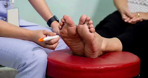
Neuropathy in diabetes has wider implications than the absence of protective pain sensation in the foot. The foot has numerous small intrinsic muscles and tendinous insertions, which help to provide stability to the digits and are delicately balanced to oppose some of the actions of the larger muscle groups within the lower leg (Kelikian and Sarrafian, 2011). Motor neuropathy results in atrophy of the intrinsic muscles of the foot (Boulton, 2007) leading to the intrinsic minus foot type: prominent long extensor tendons, cavus foot type, retracted toes or hammer toes, absence of muscle bulk and ankle equinus (Frykberg et al, 2010). The positional deformities described may have the effect of: increased forefoot pressure, plantar displacement of the first and fifth metatarsal heads; proximal interphalangeal joints of toes rubbing in shoes and poor shock absorption during the stance phase of gait. A foot with motor neuropathy and subsequent deformity is often difficult to accommodate in a ‘normal’ shoe and may not even fit in a bespoke shoe. In elective surgical practice, patients without sensory loss but with bony deformity, for example, bunion and hammer toes, desire to wear a ‘normal’ shoe without experiencing pain. This is a driver for many patients to seek major surgical intervention, such as corrective osteotomy. In comparison, insensate patients often present with similar foot deformity and foot ulceration.
Podiatric surgery
Podiatric surgery is the surgical management of the bones, joints and soft tissues of the foot and associated structures. The practice of podiatric surgery is annotated with the Health & Care Professions Council (HCPC) who approve the training programme (HCPC, 2020). National audit data show that, in the main, podiatric surgical practice is focused upon correcting painful bone and joint deformity via corrective osteotomy and foot reconstruction (Royal College of Podiatry, 2019).
The primary aim of elective foot surgery is to reduce pain, create a shoe-able stable foot, as well as improve mobility and quality of life. In addition to the core skills required to surgically manage the sensate, active, healthy patient population, podiatric surgeons are trained to manage the complications associated with surgery including, but not limited to, bone and joint infection. Podiatric surgeons are also trained in: radiology of the foot and ankle; interpretation of clinical investigations; prescribing; the management of the diabetic foot, vascular assessment, orthoses and lower-limb biomechanics.
The vascular complex foot clinic MDT
The vascular complex foot clinic (VCFC) is a weekly MDT clinic, held within a non-arterial centre, but one which still has access to on-site angiography. The clinic is staffed by a consultant vascular surgeon/consultant vascular nurse, vascular specialist nurses plus the recent addition of a podiatric surgeon. Out-patient referrals are received from the diabetes MDT, community advanced podiatrists and community nursing teams. In-patient referrals from admitted cases are also seen. The patient profile of the clinic consists of: patients with peripheral arterial disease (PAD) and tissue loss and patients with diabetes mellitus (DM) who may also have PAD, a complex foot infection or non-healing ulceration.
The MDT works closely with the diabetes teams to optimise patients medically, and podiatry teams to ensure appropriate offloading. Other hospital services, such as radiology, microbiology and the consultant antimicrobial pharmacists work remotely as part of the team (NHS Northern England, 2017). Within the VCFC, patients are assessed on an individual basis to establish the clinical urgency, underlying cause for ulceration and assess complexity in order to optimise healing.
Urgent management includes revascularisation using on-site angiography or working up for bypass procedures within the neighbouring arterial centre or surgical debridement/incision and drainage, performed by the vascular surgeons either within the Trust or at the local vascular hub. Many patients do not require urgent intervention but wound healing can be difficult to achieve due to residual infection and issues with altered biomechanics of the foot. Additionally, patients who did require urgent intervention are often left with a wound to heal via secondary intention, which can take weeks or even months to heal. These patients benefit from an assessment from a podiatric surgeon to ascertain if there would be advantages to debridement of devitalised tissue, additional management of residual infection, using local antibiotic delivery systems to fill dead spaces, debulking of a stump, resection of bone, surgical offloading or simple plastic techniques to achieve delayed primary closure.
Many of these patients are suitable for day-case surgery under the care of the podiatric surgery team. Podiatric surgeons predominantly perform elective surgery on a day-case basis under surgeon-administered regional anaesthetic (PASCOM-10, 2019) where this model of delivery is well-established, with over 20 years of national audit data demonstrating good outcomes and safety, and is transferable to many of the diabetic foot surgeries.
Outpatient procedures for diabetic foot disease cost 6.2 times less than the inpatient equivalent (Hicks et al, 2018). Regional or local anaesthetic block is a safer alternative to spinal or general anaesthesia, particularly for ASA grade three patients (Dawson et al, 2014). Surgery performed under local or regional anaesthetic is associated with reduced incidents of postoperative mortality, unplanned overnight stays, acute kidney injury and postoperative hypotension (Hammerschlag, 2008). Peri-operative insulin management is also simpler as patients are not required to fast prior to their procedure.
Despite this safer anaesthetic approach, diabetic foot surgery still carries a greater risk of other complications, such as sepsis, high-level amputation and death (Armstrong et al, 2006) and it is, therefore, essential that there is a ‘safety net’ in place for these cases in the event of a complication. Close MDT working with the diabetes and vascular teams is essential to ensure prompt admission should the patient deteriorate postoperatively. It is essential that patients and their carer’s are counselled about the early signs of complications and have precise and practical detail of how to seek treatment if required.
Surgical offloading and debridement
To ‘heal with steel’, there are cases where prompt surgical intervention may be the best option for the patient and there is an argument to say that to delay surgery is to deny healing. Of course, this is an oversimplification, and managing patients with diabetic foot disease is a complex area and requires an expert MDT approach. There are some cases where surgery should absolutely be avoided; the skill is to choose the best option for these complex cases at any given time in their care. Surgery is not a panacea for all DFUs but should be part of the discussion — particularly when combined promptly with successful revascularisation. It has been almost 20 years since Armstrong and Frykberg introduced the idea of classifying diabetic foot surgery (Armstrong and Frykberg, 2003): “Class I: Elective Diabetic Foot Surgery (procedures performed to treat a painful deformity in a patient without loss of protective sensation); Class II: Prophylactic (Procedure performed to reduce risk of ulceration or re-ulceration in person with loss of protective sensation but without open wound); Class III: Curative (Procedure performed to assist in healing open wound) and Class IV: Emergent (Procedure performed to limit progression of acute infection).”
The classification system was later validated (Armstrong, 2003) and linked to a subsequent risk of realteration and high-level amputation, unsurprisingly, those undergoing curative and emergent surgery are at a greater risk of more severe complications.
Surgical intervention should be considered on a stage II: prophylactic and stage III: curative level for suitable patients with sensory neuropathy and diabetes. Patients with significant PAD, low Toe Brachial Pressure Index (TBPI) values would not typically be suitable for stage II surgery but may benefit from stage III or early stage IV: emergent surgery post-revascularisation.
There are many surgical offloading procedures that may be performed to offload the foot in diabetes; each has its own advantages disadvantages, benefits and risks. They may be performed either in isolation or combined and are independent from the stage of surgery, for example, a flexor tenotomy may be elective or curative for an apical toe ulceration. The following list of procedures is not exhaustive and there are many variations on technique which will influence success or failure:
- Lesser toe procedures: flexor tenotomy, excision arthroplasty, partial/total nail avulsion, partial or complete digital amutation
- Hallux procedures: IPJ excision arthroplasty, accessory sesamoid excision, EHL lengthening, flexor release, Keller arthroplasty
- Fifth ray resection, weil osteotomy
- Partial ray resection +/- amputation of toe
- Exostectomy of bony prominence
- Charcot reconstruction
- Calcaneal drill and fill (LADS) for chronic OM
- Achilles tendon lengthening, gastrocnemius resection.
Local antibiotic delivery systems
In cases of deformity with ulceration and bone infection, antibiotic treatment is required. Prolonged courses of systemic oral antibiotics are associated with increased incidence of antimicrobial resistance, liver and renal impairment and Clostridium Difficile (Gauland, 2011; Fleiter et al, 2014). Morley et al (2021), in a review of 137 cases of surgical offloading/debridement combined with LADS, to be a safe and effective treatment option in a combined population of DM and PAD patients The presence of PAD was directly proportional to longer healing times and incidences of non-healing, and well perfused patients responded better to this type of intervention (Morley et al, 2021).
“The solution to pollution is dilution” — Infected soft tissue and bone may be surgically resected, the wound irrigated reducing the bacterial burden and devitalised tissue, which is a nidus for infection. Local Antibiotic Deliver Systems (LADS) such as: Cerament (V & G), Stimulan and Collatamp G, may then be inserted into the deficit and closure/partial closure of the wound performed. Utilisation of such delivery systems is well established in the US. The advantage of a topical application of antibiotic impregnated material delivers high tissue concentration of antibiotic locally, eliminating or suppressing infection and potentially preserving the limb (Morley et al, 2021).
One of the disadvantages of surgical debridement of bone is that the deficit left behind may lead to subsequent deformity despite careful rebalancing of soft tissues. In non-infected cases, bone grafts are used in order to maintain normal anatomy and restore function. Bone grafts, as well as the donor sites, are at high risk of developing infection, emerging evidence points towards the use of Cerament V or Cerament G, which is an antibiotic eluting bone graft substitute consisting of 40% hydroxyapatite (HA) and 60% Calcium Sulphate (CaS) (Bonesupport, 2021). Cerament V contains vancomycin, Cerament G contains gentamycin. Cerement can be mixed under aseptic conditions in theatre and can be moulded or shaped to fill a bone void — within 15 minutes it is set and drillable. This provides options for Cerament graft positioning with temporary percutaneous fixation.
The HA in Cerament has the ability to remodel into host bone in 6–12 months, the CaS is a carrier substance for the antibiotic medium and is slowly absorbed allowing controlled elution of the antibiotic into the wound with peak vancomycin concentrations on >3,000 mg/l and concentrations above minimum inhibitory concentration (MIC) for 28 days in vivo (Bonesupport, 2021). Postoperatively dressings must be changed regularly (1–2 days) and sterile barrier creams used on the skin surrounding the incision, as the elution of the drug can macerate wound edges.
Surgical offloading, debridement of infected tissue and the use of LADS is Class III
Curative surgery is a treatment option perhaps under-utilised nationally. The increasing use of LADS allows increased confidence to attempt primary closure following debridement or to close wounds earlier, thus reducing healing times. The following case study is patient 21 of 30 Class III cases, referred through to the podiatric surgery team from the complex vascular foot clinic. The results of larger formal clinical audit will be published in due course.
Patient case study
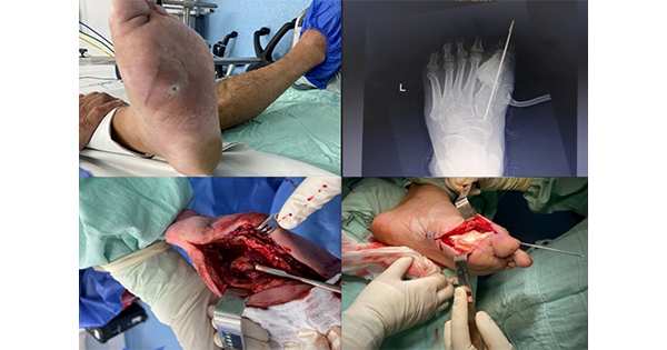
A 47-year-old male with type 2 diabetes attended the complex vascular foot clinic. He was seen by the podiatric surgeon in conjunction with the vascular nurse and consultant. He had a neuropathic diabetic foot ulceration on the plantar aspect of his fifth MTPJ (Figures 1 & 2).
Clinical examination revealed an intrinsic minus type foot with retracted toes and prominent metatarsal heads. Some reduction of muscle bulk of the plantar intrinsic musculature was identified. There was palpable, strong and regular pedal, popliteal and femoral pulses were present. He reported that, despite offloading and dressings, his ulcer had been present for 4 years. He had chronic lower back sciatic pain and was unable to tolerate a walker boot or total contact cast. Three months prior he had been admitted for six nights hospital admission for IV antibiotics for infection to the right fifth MTPJ. His BMI was 37.8, reported allergy to penicillin, all bloods were within in normal reference range other than his HbA1c 56 mmol/mol.
Pre-operative X-ray (Figures 3 & 4) revealed “cortical destruction and bony resorption noted at the volar aspect of the fifth metatarsal neck. There is associated dorsal subluxation of the fifth MTP joint. Appearances suggest active osteomyelitis. Significant soft tissue swelling at this site.”
Surgical debridement of the fifth MTPJ with Cerament bone graft substitute was discussed at length with the patient including benefits and risks, and after ensuring informed consent he was added to the waiting list under the 2-week pathway for debridement.
Procedure
Right fifth metatarsal head resection with cerement V bone graft substitute implantation under regional ankle block
Prophylactic antibiotics consisted of his current oral doxycycline 100mg BD and ciprofloxacin 500mg BD. These were continued postoperatively for 2 weeks.
The patient was positioned supine, 6 mls of 3% mepivacaine was administered by the surgeon as an ankle block. Skin prep with alcoholic betadine. The plantar ulcer was covered with a sterile Tegaderm (3M). A pneumatic ankle tourniquet was inflated to 100 MgHg above systolic pressure. Pneumatic tourniquets are not used in patients with PAD, history of revascularisation and those with vessel calcification. A stab incision to perform a tenotomy to the tendon of extensor digitorum longus was performed to aid fifth toe position, then a separate, curved dorso-lateral incision was over the right fifth MTPJ.
Delicate tissue handling and dissection straight to capsule was then incised and reflected back to reveal the infected joint. This was traced back to normal bone and resected at an oblique angle, sympathetic to the plantar and lateral surfaces of the foot. The neck of the metatarsal was dissected from soft tissues proximal to distal to the head; the bone was removed as one piece with the sinus track to the plantar ulcer. Copious saline lavage was performed. Haemostasis achieved via bipolar diathermy, blood loss was minimal. A slice of bone was then taken from the proximal end of the resection and sent to microbiology and the remainder was sent to histology.
Concomitantly, the scrubbed assistant mixed the cerement V and used a 5 ml syringe to fashion a cylinder of bone to be used for the graft — the syringe circumference being a similar size the fifth metatarsal bone. After 15 minutes, the cylinder of Cerament V bone graft was set and then shaped with a micro saw to fit the bone deficit, due to the close proximity of the plantar ulcer and likely bacterial colonisation the graft was not fixed with metalwork. Secure positioning was achieved with 2 x 2-0 absorbable sutures. Internal absorbable sutures are used sparingly in these cases and they may be a nidus for infection. The skin was closed with 3-0 prolene retention and single interrupted sutures. The skin surrounding the wound was covered with cavilon barrier cream and the wound dressed with Kendal foam, dressing gauze wool and crepe. He returned to the ward on the trolley with instructions to elevate the limb and to use a heel block shoe and crutches, with heel walking advised only. He was discharged 2 hours post-op once haemostasis assured, comfortable and routine observations were normal.
Recovery was uneventful and community nursing dressings had been arranged for the following day, with advanced podiatry input on a weekly basis. His sutures were removed 3 weeks postoperatively. The plantar ulcer had nearly healed and the incision line was healed with the exception of a small area of gapping caused by the drug elution on the incision line. He was discharged from the complex clinic to step-down advanced podiatry care. He had an appointment with the orthotist 2 days following suture removal. The X-ray review shows the implant to be maintaining the space and the fifth toe to be in a good position. The remodelling process will take a further 6–12 months.
Histology report
The histology report justified the procedure and stated: “Macroscopic examination: A part of bone measuring 20 x 15 x 13 mm. An irregular ragged soft tissue attached. The curved distal aspect, head of the bone is slightly irregular.
Microscopic Examination: Sections show focal surface ulceration with organising inflammatory slough. There is underlying active chronic inflammation extending to the subcutaneous tissues, also causing extensive bony destruction which are replaced and interspersed with areas of healing granulation-type tissue and prominent fibrosis. The features are in keeping with acute osteomyelitis. There is no evidence of atypia or neoplasia. Summary: Right fifth metatarsal head osteomyelitis.”
Microbiology culture and sensitivity
Given the clinical appearance of the wound oral antibiotics were stopped 2/52 postoperatively. The microbiology report states: “Sample bone, Corynebacterium striatum isolated, enterococcus faecalis isolated sensitive to Amoxicillin (penicillin allergic).”
Conclusion
The case study shows the curative benefits of relatively simple surgical intervention which facilitated healing of a patient with a chronic non-healing diabetic foot ulceration within a matter of weeks. It is acknowledged that in-depth vascular assessment and, if required, intervention remains a key component to ensure patient safety for surgical offloading/debridement cases. This is alongside meticulous attention to the patients’ medical management. Intervening early in the controlled theatre environment is preferable to debridement of grossly infected cases where there has been unnecessary delay or failure of prolonged conservative care. One of the skills of the experienced surgeon is, of course, knowing when not to operate. However, there is an important role for surgical intervention, established use of LADS and early closure in the US for suitable patients. This must be supported via MDT working either face-to-face — in clinic, or virtually by the expertise other medical and surgical specialties.
There are a small number of established podiatric surgery teams nationally, where they provide surgical intervention for patients with complex DFUs and they are already well-integrated within diabetes and vascular teams. Such entrenched and experienced teams are well placed to offer management of acute and chronic complex foot problems, ranging from incision and drainage of the ‘red hot’ foot to beaming for Charcot foot reconstruction. However, there is an appetite and the appropriate skill set amongst other podiatric surgeons to grow services and to improve patient care. There are opportunities to further formalise training, perhaps jointly with the vascular society and diabetes colleagues.


