Infection of the foot, usually as a consequence of skin ulceration, remains one of the major final pathways to lower extremity amputations in patients with diabetes (Apelqvist et al, 2000).
Although a multidisciplinary and aggressive approach lowers the amputation rate, limb loss due to infection remains a major problem (Larsson et al, 1995). Usually the extent of the infection is underestimated and a concomitant osteitis can be demonstrated in many cases (Lipsky, 1999).
Chronic osteomyelitis in people with diabetes is considered to have a better outcome than deep infections of the diabetic foot (Eneroth et al, 1997). The treatment of chronic osteomyelitis of the toe is considered to be surgical, i.e. removal of all the infected bone (Norden, 1999). Most reports mention an amputation of the toe or ray amputation in combination with culture-guided antibiotic treatment. In many retrospective studies, however, the results are disappointing with primary and secondary failure rates of up to 70% (Nehler et al, 1999; Murdoch et al, 1997; Wong et al, 1996).
In this retrospective study, we analysed the results of the surgical treatment of chronic osteomyelitis of the toe in 47 patients with diabetes.
Methods and materials
The Twenteborg hospital is a training hospital; there are 200000 inhabitants in this rural area. The estimated incidence of diabetes mellitus is 2% of the population. The diabetic foot unit in the Twenteborg Hospital is a subdivision of the department of surgery.
Patients with diabetic foot problems are referred by general practitioners or diabetologists. Diabetes care is supported by specially trained nurses who register and visit patients at home. Therefore, a large number of the patients with diabetes in the area are screened for foot problems on a yearly basis. About 10% of our patients are referred to us from other areas of the country.
Between 1999 and 2001, 47 patients with chronic osteomyelitis of the toe were treated; 22 were female and 25 male. The mean age was 67.8 (range 42–91) years.
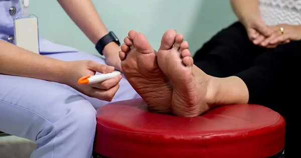
Thirty-one patients were treated with oral antidiabetics and 16 were on insulin. All patients had neuropathy, which was diagnosed using a tuning-fork (128Hz) and 10g Semmes Weinstein monofilament. All patients had absent sensation of pressure with the monofilament on four points of the foot, and had negative discrimination of vibration.
Osteomyelitis was considered present in case of signs of infection in combination with bone contact when probing, and/or the x-ray showed signs of cortical destruction (Grayson et al, 1995). If a positive culture of the probed bone was obtained, this led to the diagnosis of osteomyelitis.
None of the patients had ischaemia. Ischaemia was considered absent if pedal pulses were palpable, ankle brachial indices were above 0.8 or if the transcutaneous oxygen pressure (TcPO2) was above 30mmHg. All patients had one infected toe.
Patients were treated with antibiotics for 6 weeks. If the results of the culture were not known, a combination of clindamycin 1200mg and ciprofloxacin 1000mg daily was administered intravenously. Usually this was changed into oral administration after 2 weeks. The antibiotic regimen was adjusted according to the results of the bacterial culture.
Technique
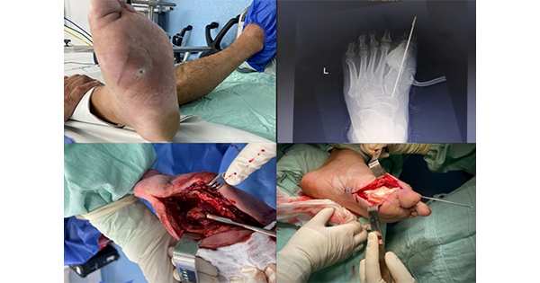
The incision was always made on the dorsum of the foot, leaving the plantar surface free of scars and avoiding weight bearing on the scar. In all cases, we avoided damage to the surrounding soft tissue, leaving as much skin as possible to achieve a tension-free closure of the wound. In all cases, the metatarsophalangeal joint was sacrificed. All patients had non-healing defects prior to surgery for more than 6 weeks. During surgery, a deep culture was obtained. In 35 of 47 (74%) patients, the wound was primarily closed without tension. In case of gross oedema or extensive redness, the wound was left open (26%). Strict bed rest was given until the wound showed a healing tendency, which we defined as a reduction in oedema and redness, and the development of healthy granulation tissue.
Open wounds were treated with an offloading cast. After wound closure, all patients received custom-made shoes. The wound was considered completely healed when there was 100% epithelialisation i.e. intact skin. Outclinic patients with incomplete wound healing were followed up on a weekly basis. Patients with complete healing were seen at 4–6 week intervals during the first year.
Results
A total of 37 of 47 (79%) patients had complete wound healing after primary surgery (Table 1). Seven of 47 patients (15%) needed a secondary intervention. Three of these seven patients had developed deep foot abscesses, which required drainage. Four patients showed persistent bone infection; three of them underwent a transmetatarsal amputation at a higher level. One patient needed a forefoot amputation. These patients eventually healed with a functional leg and ankle joint.
There were three failures of treatment (6%). Two patients eventually underwent a
below knee amputation because of sepsis and ischaemia. One patient died within 30 days after the operation due to cardiac failure (postoperative mortality was 2%). The mean time to wound healing was 106 (range 14–448) days. The mean hospital stay was 28 (range 4–128) days. The patients that were re-operated had a longer healing time of 230 (range 105–448). The mean hospital stay of the re-operated patients was 62 (range 15–128). Mean follow-up was 22 (range 12–45) months. No recurrences were seen during follow up.
Discussion
Definitive prove of osteomyelitis is a biopsy of the bone. We did not use this method for logistic reasons.
A total of 37 of 47 (79%) patients were cured after the first operation. This goal was not reached in ten patients. Seven patients healed after a second operation and a functional lower leg and ankle joint were preserved. Eventually 44 of 47 (94%) patients were cured. As all patients had antibiotics, the role of surgery alone is not clearly proven.
This study showed good results of surgery in combination with a well-selected antibiotic therapy. There are only few studies reporting the results of surgical treatment in osteomyelitis. Due to different selection criteria, comparison to our study is difficult and limited.
Nehler et al (1999) operated on 92 patients with diabetes and performed 97 amputations of the toe in patients with a forefoot sepsis. Complete healing was achieved in 34%, while in 36% the infection persisted. Twenty patients needed a below knee amputation and two patients underwent a Symes amputation.
Murdoch et al (1997) evaluated the prevalence of reamputation following resection of the great toe and first ray amputation in adults with diabetes. Sixty percent needed a second operation and 17% underwent a below knee amputation.
Wong et al (1996) described the results of 54 amputations in diabetic foot infections. Failure of surgery was described in 22 cases and the majority of these failures occurred when junior officers had operated, which stresses the importance of a good surgical technique.
Mean time to wound healing in our study was 106 (range 14–448) days. Sixteen of these patients (34%) had a healing time longer then 90 days. It is unclear why the healing time in these patients is prolonged. Yeager et al (1998) did a study to identify predictors of outcome of forefoot surgery for ulceration and gangrene in patients with diabetes. Success of revascularisation was considered as the most important predictor of initial healing and freedom of major amputation. In our study, a TcPO2 measurement of more than 30mmHg was our cut off point for ischaemia. According to literature, this value should lead to healing of wounds in 86–91% of patients (Bunt et al, 1996; Ballard JL, et al 1995).
According to the International Consensus on the Diabetic Foot (2003) a TcPO2 in the range of 30–60 in combination with intermittent claudication is considered as grade 2 peripheral arterial disease. Although these people did not complain of intermittent claudication, there might be silent ischaemia present.
Another cause of prolonged healing is persistence of infection. Surgical resection of infected areas by an experienced surgeon, with resection down to living bone, is of critical importance (Norden CW 1999). In our study, the re-operated patients all had persistence of infection and a prolonged healing time.
Although the results of conservative (medical) treatment of chronic osteomyelitis in the diabetic foot are satisfactory, the results are difficult to compare with the outcome of this study (Nix et al, 1987; Lipsky et al, 1991; Gentry et al, 1995; Senneville et al, 2001)
Selection criteria for the treatment of osteomyelitis varies between studies, and antibiotic regimen and duration of treatment differ. Some studies mention debridement but fail to specify the procedure.
In our opinion, the high percentage of healed patients in our series was achieved due to the multidisciplinary approach in each patient. The multidisciplinary foot team is supported by a group of dedicated nurses, who work strictly according to a protocol, taking young medical officers to a higher level. After discharge, all patients are checked on a regular basis in the outpatient clinic. If necessary offloading measures are taken, for instance the MaBal shoe (Hissink et al, 2000). Due to this, no re-ulcerations in the scar were reported during follow up.
Conclusion
The high healing rate of 94% as demonstrated in this series is comparable or might well be superior to alternative methods of treatment, according to the data obtained from literature.
The prolonged healing time in 34% of the patients seems to be due to persistence of infection and or silent ischaemia.
Prospective randomised trials are necessary to offer more defined guidelines of treatment for this medically, socially and economically difficult problem.


