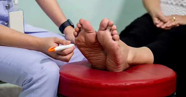
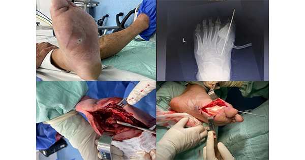
Despite improvements in the prevention and treatment of diabetes, its incidence has continued to increase (International Diabetes Federation, 2015). Diabetic foot syndrome is a challenging complication in terms of amputation risk, impact on quality of life and health-related costs (Siersma et al, 2013). The increasing prevalence of diabetic foot ulcers (DFUs) and their tendency to recur necessitates effective foot management (Pickwell et al, 2013); however, patient awareness of foot care and professional knowledge of diagnosis and management remains poor (Lipsky et al, 2015).
Acute diabetic foot infection is a rapidly evolving condition that, if not adequately treated, can quickly lead to major amputation (Faglia et al, 2006). While the pathogenetic mechanisms have been well explained, the evidence supporting treatment is far from adequate and gold standard therapeutic options are required (Aragón-Sánchez et al, 2008; Armstrong et al, 2015). International guidelines recommend prompt, aggressive surgical management with the removal of wet gangrene and drainage and debridement of abscesses or fasciitis (Peters et al, 2020). If infection is extensive or conditions are inadequately controlled by surgery, wounds can be left open to heal by secondary intent; this increases the risk of re-infection, which increases healing time (Schaper et al, 2016). Secondary wound closure by skin graft, tissue replacement or other method increases healing and reduces amputation when compared to standard care (Aiello et al, 2014); however, its use in practice is hampered by:
- A lack of strong evidence supporting effectiveness and long-term efficacy (García-Morales et al, 2012)
- High treatment costs
- The perception it is a second-line option in addition to standard treatment and needs to be embedded within a thorough multidisciplinary approach (Santema et al, 2016).
In comparison, collagen and collagen-derived metabolic components have a clear role in wound closure (Garwood et al, 2015). In normal physiology, they control many cellular functions related to healing, such as cell shape, differentiation, migration and protein synthesis (Gilligan et al, 2015). They exert local haemostatic and chemotactic stimuli while supplying a structural scaffold upon which healthy neo-tissue can develop (Shilo et al, 2013). Research to date has focused on type I collagen, a heterotrimeric protein composed of two polypeptide chains encoded by COL1A1 and COL1A2 and regulated by Janus kinase-controlled cytokines and signal transducers (Tabib et al, 2018). Post-translational changes activated by specific hydroxylases and glycosyl-transferases transform type I collagen into its final form (Varma et al, 2016). Its close mimicry of normal collagen allows it to act as a fibroblast attractant, eliciting further collagen deposition at the treatment site and triggering positive feedback that accelerates regeneration and repair (Shoseyov et al, 2014).
Sources of collagen
Collagen-derived products generally come from cadavers or animal sources (Lane et al, 2018).
Products derived from animal sources result in cellular and humoral immune responses in about 10% of patients that are self-limiting within few months but have been associated with impaired collagen function (Benders et al, 2013). Their use is contraindicated in patients with hypersensitivity to collagen or bovine products. The need for a skin test, the results of which are generally available in 2–4 weeks, strongly limits their clinical application (Ganesan et al, 2018). Bovine products present a risk of prion transmission, so regulatory authorities have limited their use in countries where bovine spongiform encephalopathy has been reported (Moody et al, 2001).
Human-derived collagen reflects the clinical characteristics of the source, as age, ethnicity, environmental setting and genotypes modify its characteristics (Gabler et al, 2018). Increasing age results in molecular crosslinking, reducing elasticity and acid solubility and increasing resistance to collagenases (Luong et al, 2019). Furthermore, extraction of collagen and its derivatives introduces intra-molecular crosslinks that reduce solubility and scaffold-assembling capacity (Oryan et al, 2015).
Plant-made pharmaceutics overcome these obstacles (Baroni Edo et al, 2012). The tobacco plant is ideal for bioengineering as it is easy to grow under controlled conditions and its high leaf mass allows for large-scale protein extraction. Tobacco plants are engineered to express five different human genes: two encoding type I collagen subunits and three encoding human enzymes critical for post-translational modification of type 1 pro-collagen. Pro-collagen is extracted from tobacco leaves and purified to produce pure human recombinant type 1 collagen (Fisher et al, 2005). This molecule is a pure heterotrimeric collagen free from high-molecular-weight contaminants and rich in hydroxylated prolines and lysines, similar to human collagen (Sierra-Sánchez et al, 2020).
The collagen assembles itself in a three-dimensional structure that, in contrast with human- or animal-derived collagen, maintains a wide contact area with integrin-binding sites and has a high water-retaining capacity (Table 1) (Shoseyov et al, 2013). It has similar scaffolding properties to the human wild type and is able to trigger deposition of human collagen itself (Bhowmick et al, 2015).
Recombinant type I human collagen gel
This is an advanced wound care treatment produced by genetically-engineered tobacco plants. It is available in two formulations: a wound dressing matrix and a gel. The gel has been created to manage tunnelled and/or undermined wounds. The formulation maximises contact with the wound bed and surrounding tissues, enabling scaffolds to develop across the wound bed (Sharma et al, 2017). Its homogeneity means that the gel can produce strongly ordered fibres and membranes that orient molecules in the same direction, resulting in new tissue with better tensile strength and greater elasticity (Willard et al, 2013).
Aim
This study aimed to evaluate the safety and efficacy of recombinant type 1 human collagen from tobacco plants in the management of post surgical diabetic foot wounds left to heal by secondary intention.
Method
Participants
Adults with diabetes admitted to the inpatient clinic were assessed for inclusion in this study between March and July 2017. Patients with lower limb complications who underwent a surgical procedure and whose wounds were left open to heal by secondary intent were eligible to participate. Patients were excluded when:
- Local clinical infection was present after surgical debridement or systemic infection was present and uncontrolled by antibiotics
- Critical limb ischaemia (CLI) was present and not amenable to revascularisation
- The wound was closed by primary intention
- Negative pressure wound therapy was required
- HbA1c >108 mmolmol (12%)
- Disease was potentially interfering with tropism and skin repair
- Active neoplasia was present
- There was hypersensitivity to treatment
- Life expectancy was <1 year.
Participants were randomised to standard treatment consisting of local debridement as described by international guidelines or standard treatment plus recombinant type I human collagen. The collagen was applied in the operating room after surgical debridement and covered with a polyurethane film dressing.
On admission, patients provided formal consent for their data to be added to the database and used in a non-nominal aggregate form. Patients who agreed to participate signed an informed consent form. The study protocol was approved by the local ethics committee.
Protocol
On admission, patients were checked for signs of infection, underwent vascular assessment with duplex ultrasound and had their DFUs assessed. CLI was treated by percutaneous angioplasty or arterial bypass grafting, when feasible, followed by ultrasound to determine whether revascularisation had been successful. If wet gangrene, pus or extensive infected necrosis was present, wounds were debrided before revascularisation; otherwise, debridement and minor amputation were performed after revascularisation (Bakker et al, 2016).
Each DFU was graded using the University of Texas classification and its location recorded (Oyibo et al, 2001). DFUs were routinely swabbed during the first clinical evaluation and any empirical antibiotics given tailored according to culture results.
Two days after surgery, the external bandage was removed to check the collagen and the external polyurethane film. In the absence of complications, their risk factors were assessed and they were discharged with a fixed offloading system (Optima Diab™) in place.
After discharge,patients were followed up weekly until complete re-epithelialisation. Wound data were collected using the Visitrak measurement system. Data on complete healing, recurrence, and presence and severity of adverse events were collected using Upodi.
Outcomes
The primary endpoint was the rate of healing. The secondary endpoints were:
- Healing time — the time between surgery and complete re-epithelialisation of the target lesion confirmed on 2 consecutive weeks
- Reduction in wound area
- DFU recurrence.
Statistical analysis
Quantitative variables were expressed as mean, median and standard deviation; qualitative variables as frequencies and percentages. Categorical data were compared using chi-squared and Fisher’s exact test. Continuous variables were examined using student’s t-test. Statistical analysis was performed using SAS software (SAS Institute, Cary, NC); p<0.05 was considered statistically significant. The association between healing and the main clinical characteristics was assessed by Cox proportional regression. Hazard ratios (HRs) were reported with their 95% confidence intervals.
Results
Between March and July 2017, 186 patients were admitted: 12 with glycometabolic decompensation, hypoglycaemia or other conditions not associated with lower-limb complications and 174 with diabetic foot-related problems. Of the diabetic foot patients, 33 did not require surgery. Of the 141 that underwent diabetic foot surgery, 98 had wounds that were sutured during the procedure to promote healing by primary intention. The 43 remaining patients’ wounds were left open to heal by secondary intention. Of these, 24 patients met the inclusion criteria and were enrolled in the study. The selection process is given in Figure 1.
Twelve patients were randomly assigned to collagen plus standard treatment (Group A) and 12 to standard treatment alone (Group B). There were no significant between-group differences in characteristics (Table 2) or surgeries performed (Table 3).
Four participants (16.7%) presented with CLI and underwent percutaneous transluminal angioplasty before any surgical procedures. All procedures were successful. One of the revascularised patients was randomised to Group A and the other three to Group B. No differences were observed in transcutaneous oxygen tension at the time of treatment (52±8 mmHg in Group A; 55±6 mmHg in Group B).
Post-surgery, the wounds had a mean area of 8.2±3.8 cm2 (range 1.2–9.7 cm2). Seventeen (70.8%) patients’ wounds completely healed during follow-up, with a mean healing time of 78±9 days. The healing rate was significantly higher in Group A (10 out of 12 patients; 83.3%) than Group B (7 out of 12 patients; 58.3%). Healing time was significantly shorter in Group A than Group B (64±4 days versus 90±11 days, p<0.02).
Seven wounds had not completely healed by the end of the study. There was a mean 63% reduction in wound size but reduction was significantly greater in Group A than Group B (78% versus 50%, p <0.02), see Figure 4.
There were no DFU recurrences or re-interventions required during follow-up. No local or systemic adverse events or hypersensitivity reactions were reported.
Univariate Cox regression analysis, see Table 5, found male sex, older age, HbA1c >64 mmol/mol (>8%) and diabetic retinopathy decreased the chances of wound healing. The application of recombinant type I human collagen treatment and baseline wound size <5 cm2 were associated with complete healing. Multivariate regression analysis confirmed that type I collagen (HR 1.78, p=0.02), older age (HR 0.82, p<0.03), glycometabolic disturbance (HR 0.78, p=0.04) and wound size <5 cm2 (HR 1.56, p<0.01) but not male sex (HR 0.92, p=0.23) or diabetic retinopathy (HR 0.82, p=0.45) had an impact on wound healing, see Table 5.
Discussion
Diabetic foot is a serious problem in Western countries. Population ageing and improvements in diabetes control have led to an increase in the prevalence of lower limb complications, which are complex and have a high risk of relapse. The creation of an individualised treatment strategy is a cornerstone for obtaining real improvement in quality of life. To reduce the impact of DFUs, careful and wide debridement of non-vital tissue should be performed to save limbs and lives as part of a treatment pathway that restores functionality of the limb in the shortest possible time. This requires the application of a wide range of therapeutic options, e.g. negative pressure wound therapy and dermal substitutes.
Collagen therapies support healthy epithelial cell migration, creating a scaffold that supports cellular development and enhances tissue regeneration. The use of collagen and other materials in the secondary closure of infected diabetic foot wounds is still debated. For many years the only collagen products available were human- or animal-derived, which come with risks associated with their molecular cross-mimicry. The development plant-derived collagen products has increased the recent use of collagen (Shilo et al, 2013). Diabetes is strongly associated with a reduced capacity to create scaffolds for fibroblast development and with chronic prolonged hyperglycaemia, which exacerbates this issue. The application of collagen products results in significant cellular regeneration and activates cellular pathways typically impaired in diabetic foot syndrome (Frykberg et al, 2016). Many collagen-containing formulations have been developed. Gels allow collagen products to fill cavities and adhere to the wound bed, preventing maceration. Many studies have explored the action of collagen gel on wound healing outcomes but this is the first randomised controlled trial on the use of recombinant human type I collagen in diabetic foot wound management.
The groups in the study had comparable clinical and demographic characteristics. There were two non-significant differences between the groups: Group B had a higher proportion of individuals with type 1 diabetes, which was not associated to a longer duration of diabetes; and Group A had a higher number of patients with previous lower-limb revascularisation. The latter could have impacted clinical outcomes due to the impact of ischaemia on wound repair; however, all the revascularisation procedures restored a good blood supply to the foot, as witnessed by near-normal levels transcutaneous oxygen tension.
This study found the use of recombinant type I collagen to be associated with shorter healing times and a higher chance of complete healing in post-surgical diabetic foot wounds. As expected, it was more effective in younger patients with a good glycometabolic control. Smaller baseline wound size was a positive prognostic factor for healing. The presence of diabetic retinopathy, as confirmed in previous trials, was associated with a poorer prognosis (Siersma et al, 2017). No adverse reactions or hypersensitivity were reported during trial and follow-up period, suggesting that the use of type I collagen is safe.
Costs associated with collagen products limit their application in clinical practice. Our findings suggest the cost of type I collagen would be offset by savings resulting from faster and higher healing rates. More rapid healing reduces direct healthcare costs (admissions, procedures and dressings) and reduces personal costs to the patient (improved quality of life, faster return to work/social activities). Despite these potential savings, the costs associated with treatment necessitate careful patient selection to maximise benefits. Clinical research should focus on identifying which patients would benefit most and the point at which application is most effective.
Limitations
This study included a very small number of patients and was limited to a single tertiary referral centre, limiting the generalisability of the findings. However, the results support the argument for a larger prospective randomised controlled trial in multiple centres and settings to better quantify the benefits of recombinant type I human collagen on DFU healing and which patients derive the greatest benefit.
Other study limitations relate to the cost and the difficulty in obtaining large amounts of recombinant type I collagen. Despite these issues, the authors felt the novelty of the product tested and its potential benefits in the management of complex DFUs justified this pilot trial. These issues will need to be addressed if this treatment is to be applied in daily clinical practice. Further studies and technical improvements are needed to overcome these limitations.
Conclusion
Recombinant type I human collagen produced by tobacco plants promotes tissue repair at a cellular level and has great potential as a treatment for diabetic foot wounds. Its addition to standard care in the management of post-surgical diabetic foot wounds resulted in faster healing by secondary intention and a higher rate of healing than standard care alone. As it is expensive, a larger, multi-centre follow-up trial is required to clearly identify those who will benefit most from treatment and to quantify its advantages over standard care.


