To complete the CPD module, click here.
In 1868, the neurologist Jean-Martin Charcot described a chronic and progressive neuroarthropathy resulting in joint destruction in patients with tabes dorsalis (Charcot, 1868). However, it was not until 1936 that the first case of neuropathic arthropathy was described in a person with diabetes (Jordan, 1936).
Today, diabetes is the most common cause of Charcot neuroarthropathy (CN) in developed countries (Sanders at al, 1993, Armstrong et al, 1997; Trepman et al, 2005; Frykberg et al, 2006; van der Ven et al, 2009). Other causes include syphilis, HIV, leprosis, syringomyelia, meningomyelocele, spina bifida, amyloid neuropathy, neuropathies secondary to alcoholism and renal dialysis, and postrenal transplant arthropathy (Sequeira, 1994; Wukich and Sung, 2009; Mabilleau and Edmonds, 2010).
CN is characterised by a progressive process of deterioration of weight-bearing joints, most commonly the foot and the ankle, and is a challenging clinical condition. It causes progressive bone deformity and osteoarticular instability, which leads to ulceration, infection and, consequently, a elevated risk of amputation (Armstrong and Peters, 2002; Rajbhandari et al, 2002; Sanders, 2008; Rogers at al, 2011; Frykberg and Bleczyk, 2012). Pathologic fractures and dislocations are associated with CN, with collapse of the architecture of the foot and progression to plantar deformity.
CN is also known to lead to a reduction in quality of life, high disability and an increased risk of mortality (Pakarinen et al, 2009). Despite its relatively low incidence (3–11.7 per 1,000 patients per year) and low prevalence rates (from 0.08% to 7.5%), CN causes significant mortality and morbidity, with mortality rates estimated to be as high as 28% (Armstrong et al, 2002; Frykberg et al, 2006; Sohn et al, 2009 Wukich and Sung, 2009).
CN is commonly misdiagnosed as sprain, deep vein thrombosis, osteomyelitis, cellulitis or rheumatoid arthritis. The earlier the correct diagnosis and the initiation of appropriate treatment, the smaller the risk of developing deformity and articular instability that can lead to ulceration, osteomyelitis and amputation (Sinha et al, 1972; Giurini et al, 1991).
This review focuses on the pathogenesis, clinical features and therapy of CN, with an emphasis on the surgical options.
Pathogenesis
It is currently believed that CN occurs because of an episode of inflammation in the foot becoming abnormally protracted as a result of underlying neuropathy. The inflammation may be triggered by a number of factors (such as loss of protective sensation, neuropathy-mediated abnormalities of distal blood flow, or loss of innervation of bone), but the resultant expression of pro-inflammatory cytokines (such as interleukin-1 beta, tumour necrosis factor alpha) leads to activation of the receptor activator of nuclear factor kappa-beta ligand (RANKL/NfkB pathway).
The nuclear transcription factor NfkB triggers the maturation of osteoclasts and these cause bone lysis (Jeffcoate et al, 2005). Such an activation of osteoclasts is part of the normal response to injury and facilitates the clearance of debris before the onset of wound repair, but the process is normally short-lived. If, however, the sensation of pain is reduced, or even absent, patients will continue to walk on the inflamed foot, and this initiates a cycle of uncontrolled inflammation with progressive osteolysis. This further weakens the pedal skeleton, making it prone to progressive fractures and dislocation (Baumhaauer et al, 2006; Uccioli et al, 2010; Kaynak et al, 2013).
It should, however, be noted that the evidence to confirm involvement of the RANKL/NfKB pathway in the pathogenesis of the Charcot foot is largely circumstantial. Mabilleau et al (2008) have suggested that a RANKL independent mechanism might be involved. There is some early interest in the osteoblast-dependent osteogenic mechanism mediated via the Wnt/beta-catenin pathway and its endogenous inhibitors, sclerostin and dickkopf-1. The Wnt/beta-catenin pathway is involved in osteoporotic disease and has been shown to be responsive to anti-sclerostin monoclonal antibodies (Becker, 2014). It is particularly interesting to note that — just like the RANKL /NfKB pathway — the Wnt/beta-catenin pathway is disordered in diabetes, and that both are implicated not only in abnormal bone breakdown, but also in parallel increases in macrovascular disease (Jeffcoate et al, 2009; Petrova and Shanahan, 2013; Gaudio et al, 2014).
Petrova and Shanahan (2013) demonstrated that the osteoclasts in people with CN are more aggressive than controls and that the inhibition of TNF-alpha is able to reverse the increased bone resorption. The more recent interpretations of this pathogenesis of CN tend to give the accidental repetitive trauma in the insensate neuropathic foot the role of the activator of a pathologically increased inflammatory cascade mediated by cytokines and actuated by the osteoclasts.
Presentation and diagnosis
CN may present with isolated inflammation of the foot (with or without discomfort), and is eventually associated with varying degrees of destruction of the architecture of the foot.
The acute phase of the disease is characterised by severe swelling, warmth and erythema of the foot, associated with bony resorption and fragmentation. This acute inflammatory phase may lead to varying degrees and patterns of bony destruction, subluxation, dislocation and deformity (Rogers et al, 2011). The distortion of the foot may lead to ulceration of the skin over areas exposed to abnormal forces and this may lead to infection. The bones and joints most often affected are those of the midfoot and hindfoot, with fractures accompanying medial dislocation of the second to fifth tarsometatarsal joints and downward dislocation of the talonavicular joint. Any of these may lead to the classical infero-medial bulging of the foot with loss of the plantar arch, often referred to as “rocker bottom foot”. Other cases of what appear to be same process are much more limited in their extent, such as isolated fracture of one or two metatarsals (Jeffcoate, 2015).
The majority of people who present with an active Charcot foot will recall seemingly minor trauma that they relate to the onset of symptoms. Sometimes this might have been very recent, but sometimes the delay is weeks or months. One survey reported that these episodes include minor accidental trauma (recalled in 36% of all presenting cases), preceding foot ulceration (35%), local surgery (12%), and osteomyelitis (7%) (Game et al, 2012).
In the acute active stage, the patient typically presents with unilateral-dependent erythema, oedema and increased skin temperature, generally 2˚C warmer more than the contralateral foot and ankle (Rogers and Bevilacqua, 2008). In the acute active stage, the disease is often mistaken for cellulitis. If the skin is intact, these findings are pathognomonic of CN. In some patients, the diagnosis may be complicated by concomitant ulceration that raises the possibility of osteomyelitis. Pain may be absent due to the presence of peripheral neuropathy; pain actually occurs in more than 75% of patients, but normally it is less severe than expected in the presence of the significant clinical and radiological findings (Petrova et al, 2004; Baglioni et al, 2012).
The main barrier to a prompt diagnosis of CN is the failure of the clinician to consider the possibility. Most frequently, affected people are examined by non-specialists in primary care and emergency departments where the most common causes of this symptomatology are sprains, cellulitis, venous thrombosis and gout. It follows that many people with an active Charcot foot are investigated, and even treated, for one of these conditions before the correct diagnosis is made (Pinzur, 2007a). The delay in diagnosis is likely to result in a worsening of the disease and may increase the risk of eventual limb loss (Pakarinen et al, 2009; Chantelau and Ricther, 2013).
The association of the clinical presentation with radiological evidence of fracture and/or dislocation is usually sufficient to make the diagnosis and to start treatment. If the X-rays appear normal, it is essential that magnetic resonance imaging (MRI) is requested as soon as possible to identify inflammation of soft tissue (which is not diagnostic) and bone marrow (which is strongly suggestive of active Charcot foot). If the MRI is negative, it is usually accepted that a diagnosis of Charcot foot is unlikely, although specialist teams will occasionally encounter cases in which MRI later becomes abnormal (Jeffcoate, 2015).
Classification
Several systems have been proposed to classify CN. Among these, two classifications are widely quoted. The Eichenholtz Classification (Table 1) — especially in the modified version, which includes a stage 0 — is essentially a summary of the theoretical stages through which the CN is thought to progress: from inflammation without skeletal damage, to skeletal damage and possibly to resolution (Eichenholtz, 1966).
The Sanders and Frykberg classification (Table 2) stratifies the disease on the basis of the affected joints. It identifies five patterns of damage in the foot and ankle associated with different anatomical patterns (Sanders, 1991, 2008; Frykberg et al, 2012). The location at the Lisfranc joint and at the ankle/subtalar joints are the most severe structural deformities and instability in these joints is associated with a major amputation risk. Type V is rare, but it may be correlated with an avulsion injury or isolated pathologic fracture of the posterior calcaneal tuberosity.
The most recent classification is the one proposed by Chantelau and Gruztner (Table 3). This is based on MRI, dividing affected feet into active or inactive arthropathy, as well as whether they are associated with full thickness cortical fractures (Chantelau and Grutzner, 2014)
Clinical management
Immediate referral to a specialist clinic or a multidisciplinary foot clinic for management is indicated in any case of suspected CN (Chantelau, 2005). The primary goals of treatment are structural stabilisation of the foot and ankle, with maintenance of a plantigrade, stable foot, able to fit into a shoe, in order to prevent recurrence and reulceration. Immediate immobilisation and offloading of the foot and ankle remains the cornerstone therapy during the active stage of CN.
The medical management of CN has evolved over the past few years, with an emphasis on drugs that regulate osteoclatic activity because osteopenia has been shown to be more prevalent in patients with diabetic neuropathy and CN (Petrova et al, 2004). Bisphosphonates have been proposed as a treatment since they slow down osteoclastogenesis (Selby et al, 1994). However, a recent systematic review concluded that there is not enough evidence to support the use of bisphosphonates in the management of CN (Richard et al, 2012). Other methods, such as intranasal calcitonin and bone stimulation, have been studied, but randomised controlled trials are still lacking (Bem et al, 2006).
Offloading
Immediate immobilisation and offloading of the foot and ankle remains the cornerstone of therapy during the acute stage of CN (Stefansky and Rosenblum, 2005; van der Ven et al, 2009; Wukich and Sung, 2009). The goal of conservative treatment is to interrupt the destruction process and to maintain adequate foot and ankle alignment and a plantigrade position compatible with stability and walking.
The International Consensus on Charcot Foot recommends the use of a moulded below-knee fibreglass cast, which is easy to apply and strong enough to allow weight-bearing once it has set (Bakker et al, 2016). Non-removable casts should be changed within 1 week because if there is a prompt reduction in local inflammation, they rapidly become loose. Thereafter, casts need to be replaced every 1–3 weeks. Frequent replacement gives an opportunity for the foot to be checked — not least because the cast may cause abrasions of which the patient may be unaware because of the underlying neuropathy.
Non-weightbearing on the affected joint should be prescribed until the resolution of the destructive phase, in order to interrupt the cycle of repetitive trauma and to minimise fracture and debilitating deformities, until the clinical signs of acute inflammation completely regress, which will take 2–6 months (Pinzur et al, 2006; de Souza, 2008).
When to consider surgery
Surgical procedures are recommended when all conservative treatments fail to prevent ulcerations (Pinzur et al, 2006). The goals of surgical treatment are to preserve functional activity, to restore stability and alignment so that appropriate footwear or bracing is possible, and to prevent amputation.
The indications for CN surgery are: exostosis with high-risk of ulceration despite an optimal orthotic treatment, severe articular instability, pain associated with misalignment and relapsing wounds, associated osteomyelitis, and selected acute fractures (Burns and Wukich, 2008).
A complete workup and optimisation is important to achieve a successful surgical outcome and to stratify the risk of complication.
The preoperative evaluation should include knowledge of patient’s status, a cardiologic assessment, possibly integrated with a stress test to highlight an inducible ischaemic heart failure (Wukich et al, 2016), the evaluation of the quality of soft tissue, and an appropriate vascular assessment, and in case of critical limb ischaemia, a peripheral percutaneous or surgical revascularisation (Dalla Paola et al, 2016).
Surgical planning must take into account the clinical condition, with careful consideration of the exostoses and their location: the patient should be clinically evaluated in offloading and weight- bearing state. The standard X-ray will evaluate the deformity that is evident on the sagittal plane (Figure 1), assessing the lateral tarsal-first metatatarsal angle (Meary’s angle), cuboid height, medial column height, calcaneal fifth metatarsal angle, lateral tibio-talar angle, and the transverse plane (Wukich et al, 2014).
The appropriate timing for surgery is usually considered at Eichenholtz stage II–III, because in the active phase the risk of failure of stabilisation is considered high due to the active inflammatory status and to the osteoarticular destruction. However, some authors report successful arthrodesis and open reduction and internal or external fixation during the developmental stage: Simon et al (2000) reported the use of early arthrodesis for Eichenholtz stage I, noting the likelihood if anatomical reduction, clinical union, and stability with or without increased risk of complications.
There are no robust data on which to base a choice between surgical and non-surgical treatment during the active phase of CN. There are currently no data showing the opportunity to move surgery to earlier stages of the disease. The prevailing opinion is defined in the German-Austrian consensus statement that a deformed, but plantigrade, foot capable of full weight-bearing is not a candidate for surgery, and surgery should be considered only for cases with joint dislocation and significant instability because reduction and retention by means of casting is ineffective (Koller at al, 2011).
Surgical treatment options
Surgical options range from simple decompressive exostectomy to more extensive realignment and arthrodesis of the foot and ankle with internal or external fixation. Bone correction is achieved with exostectomy, osteotomies and/or arthrodesis. Sometimes, in very complex deformities, a combination of the three approaches is required.
Exostectomy
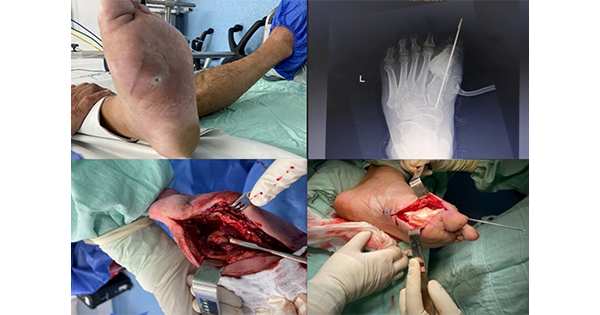
Simple exostectomy is indicated for stable midfoot CN in order to prevent primary recurrent ulcerations and to relieve shoe-fitting problems (Brodsky and Rouse 1993; Catanzariti et al, 2000; Simon et al, 2000; Mueller et al, 2003; Laurinaviciene et al, 2008). It consists of surgical removal of the bony prominence from the apex of the rocker bottom deformity of the foot (Figure 2). A concomitant percutaneous lengthening of the tendon or gastrocnemius recession is often required to achieve a plantigrade foot and to decrease the chances of ulcer recurrence (Catanzariti et al, 2000; Mueller et al, 2003). One potential complication from exostectomy is an iatrogenic midfoot instability from an aggressive resection or failure to recognise potential preoperative instability. Exostectomy is most effectively used when tarsometatarsal joints are involved (Shen and Wukich, 2013).
Realignment arthrodesis with internal fixation
The major goal of reconstructive arthrodesis is to restore the stability and alignment of the foot and ankle, so that the prescription footwear can be worn. It involves resection of nonviable bone with reduction of the deformity followed by the stabilisation and/or arthrodesis of multiple tarsal and/or ankle joint. This procedure is indicated for failed exostectomy, gross instability, severe fixed deformity and acute dislocation. The surgical approach depends on the severity and location of deformity, and the level of instability.
The internal fixation systems for CN with involvement of the foot or ankle include intramedullary implants (screws, cannulated screws, nails, rods) or extramedullary implants like locking or nonlocking plates or fixed angle plates. Recently, solid or cannulated intramedullary screws (i.e. midfoot beaming) have been used in CN midfoot fusion. The advantages described are restoration of anatomic alignment and fixation beyond the localisation of deformity (Lamm et al, 2012).
Methods of stabilisation for midfoot CN include multiple screw fixation, staples, intramedullary screws, and compression plating and screw constructs (Papa et al, 1993; Sammarco et al, 2010; Lowery et al, 2012; Pope et al, 2013).
The use of a plate on plantar aspect of the medial column of the midfoot has been advocated to enhance the rigidity of an arthrodesis of the midfoot (Schon and Marks, 1995).
Open reduction and arthrodesis with use of multiple axially placed intramedullary screws provided reliable construct to achieve and maintain correction of deformity (Sammarco et al, 2010).
The CN localisation to the ankle corresponds to the earliest indication for surgery: ankle and hindfoot CN can be successfully managed with a retrograde intramedullary revision ankle nail (Pope et al, 2013).
Moreover, a displaced unstable ankle fracture is an indication for immediate open reduction and internal fixation (Lowery et al, 2012).
Long-term immobilisation is crucial for achieving union; generally, the duration of immobilisation after arthrodesis for patients with CN is twice as long compared to nondiabetic patients. Postoperative treatment consists in casting and total non-weightbearing for at least 12–18 weeks and further partial weight bearing for 3–6 months until consolidation has been achieved (Idusuyi, 2015).
Realignment arthrodesis with external fixation
External fixation (Figure 3) has gained popularity as a less invasive treatment of CN (Cooper, 2002; Farber et al, 2002; Zarutsky et al, 2005; Roukis and Zgonis, 2006; Zgonis et al, 2006; Pinzur, 2007b; Dalla Paola et al, 2009). External fixation systems have different features and their use varies according to the situation. There are several categories of external fixators — static, dynamic or for offloading stabilisation. Many papers confirm the possible positive approach of external fixation on different pathophysiological aspects of CN, including decreased bone mineral density, bone loss, osteomyelitis, non-union, peripheral vascular disease, and compromised soft tissue coverage of the surgical site (Cooper, 2002; Jolly et al, 2003; Wang, 2003; Lamm and Paley, 2006; Pinzur, 2007b; Matsumoto and Parekh, 2015; Lee et al, 2016).
Small wire circular fixators may be used when there is an open wound or when bone loss preludes satisfactory open reduction. A study by Dalla Paola et al (2009) showed that external fixation is a valid alternative to major amputation in selected patients.
Internal or external fixation?
Choosing the fixation method (internal/external or hybrid) depends on a complex evaluation that considers the bone quality, soft tissue condition, the presence and entity of fractures and/or dislocation, history of previous surgery, walking ability, comorbidities and degree of obesity. Internal and external fixation techniques have been described for midfoot Charcot deformities, but the lack of comparative studies makes it difficult to advocate one technique over the other. Recently only one systematic review has compared the short term outcomes (mean follow-up 35.7 months) of internal versus external fixation for Charcot midfoot neuroarthropathy, although it is based on non-randomised, non-controlled, and predominantly retrospective studies (Lee et al, 2016). This systematic review, even if limited by the quality of included studies, justifies the use of external fixation as an alternative to internal fixation. Although internal fixation may decrease the risk of non-union and increase return to functional ambulation, it may result in significantly more complications than external fixation. External fixation was associated with a higher rate of ulceration, but resulted in fewer cases with any complication, including a decreased risk of extremity amputation, deep infection, wound healing problems, peri- or intraoperative fractures, and the needs for unplanned further surgery.
Surgical management of Charcot foot complicated with osteomyelitis
The therapeutic plan for CN with concurrent osteomyelitis (OM) is extremely complex and often lengthy. This is due to the need for a sequence of multiple surgical procedures, prolonged antibiotic therapy, extended periods of non-weightbearing and immobilisation, and use of specific technologies such as negative pressure wound therapy, engineered tissues, external fixation. These therapeutic plans and protocols should be managed by multidisciplinary teams specifically trained on the subject because of the extreme complexity of these patients and their serious comorbidities (Dalla Paola, 2014). However, the biggest issue is still the comparison between reconstructive treatment and primary amputation (Dalla Paola, 2014).
Once patients develop osteomyelitis, the decision-making algorithm becomes complicated. A significant percentage of these patients will require amputation, often after failed reconstruction attempts and multiple courses of antibiotic therapy (Sohn et al, 2010).
Many experts currently believe that deformity correction in patients with CN greatly improves their quality of life, fosters greater walking independence, and improves longevity. Instead, detractors suggest that surgery is not justified, given the cost of care and the risk associated with its complexity (Sohn et al, 2010; Dalla Paola, 2014).
Data from a recent study by Gil et al (2013)suggest that the cost of care for successful transtibial amputation may be very similar to the cost of limb salvage, at least during the first year.
Another significant factor to consider is that these patients often have an elevated body mass index and are unlikely to achieve independent ambulation following below-knee amputation and prosthetic limb fitting, thus justifying conservative reconstructive surgical procedures.
Evidence base
Unfortunately, the evidence available for CN surgery is scarce and mostly based on retrospective case series. As a consequence, surgical algorithms for the treatment of CN of the foot are based almost entirely on level IV or V evidence. From the current limited and relatively weak published data (uncontrolled retrospective case series and case reports), it is possible to make the following points:
- There is some inconclusive evidence that surgery performed during the acute phase of CN is useful
- Exostectomy is useful to relieve bony pressure that cannot be accommodated by orthotics and prosthetic means
- Prophylactic surgery (Achilles tendon or gastrocnemius muscle lengthening) reduces forefoot overload and improves the alignment of the ankle and the hindfoot to the midfoot and the forefoot
- Arthrodesis in cases of instability, pain or recurrent ulcerations is indicated, with good outcomes, despite a relatively high rate of incomplete bony union (Lowery et al, 2012).
Conclusion
Early recognition and intervention can limit the deformity associated with Charcot foot. Aggressive conservative management should be initiated early in the treatment plan in an effort to minimise the devastating effects often seen with this condition. Any delay in therapy can result in severe foot and ankle deformity, in which traditional nonsurgical methods alone may be inadequate. These deformities may lead to ulcerations and ultimately progress to amputation. Surgical correction and stabilisation is an effective method to prevent further deformity and ulcer recurrence, and if it is performed in appropriate setting, according to the right indications, CN reconstruction is a better alternative to lower-limb amputation.
There currently is no consensus on the optimal method of surgical correction, as each technique has strengths and weaknesses. There is inconclusive evidence concerning timing of treatment and use of different fixation methods. Prospective series and randomised studies, even if difficult to perform, are necessary to support and strengthen current practice.
Although surgeons who reconstruct Charcot deformities may feel the surgery is beneficial, so far no study has been done comparing surgical correction to non-operative treatment or amputation.


