
Diabetes is one of the main global public health issues and is a substantial burden on public health and socio-economic development (Lin et al, 2022). The International Diabetes Federation (2021) estimates that approximately 537 million adults (20–79 years) are living with diabetes in 2021 and the total number of people living with diabetes is projected to rise to 643 million by 2030 and 783 million by 2045. In 2016, the World Health Organization stated that the rate of diabetes in China is “explosive”’, and noted that “almost 10% of all adults in China — about 110 million people — currently live with diabetes” (Wu, 2016).
The increase in the pervasiveness of diabetes has led to an increase in the associated complications of the disease, among which foot ulcers and lower extremity amputations are notable (Atosona and Larbie, 2019). Diabetic foot ulcers (DFU) have been described as the main complications of diabetes and a foremost source of morbidity and mortality among people with diabetes (Abdissa et al, 2019; Atosona and Larbie 2019).
The International Working Group on the Diabetic Foot (IWDGF) states that, globally, every 20 seconds a leg is amputated due to diabetes and the lifetime incidence of DFU in people with diabetes is 15–20%, with the possibility of recurrence within the first year being 30–40% (Van Netten et al, 2016; Armstrong et al, 2017). It is estimated that 12.9 million to 49.0 million people worldwide have a history of foot ulceration (Armstrong et al, 2017). Moreover, the risk of death at five years for a patient with a DFU is 2.5 times as high as the risk for a patient with diabetes who does not have a foot ulcer, and mortality after diabetes-related amputation surpasses 70% at 5 years (Lavery et al, 2010; Walsh et al, 2016).
However, a DFU may heal in many patients with the application of appropriate therapy, including surgical debridement, offloading, prevention and management infection, and, where necessary, vascular reconstruction (Bus et al, 2016; Hinchliffe et al, 2016).
The IWGDF practical guidelines describe the basic principles of prevention and management of diabetic foot disease and consist of evidence-based principles of prevention of foot ulcers, offloading, diagnosis, prognosis and management of peripheral artery disease in patients with a foot ulcer and diabetes, diagnosis and treatment of foot infection, interventions to enhance healing, and classification of diabetic foot ulcers (Schaper et al, 2019).
It is interesting to note that the IWGDF recommends the consideration of the use of sucrose-octasulfate impregnated dressings as an adjunctive treatment, in addition to best standard of care, in non-infected, neuro-ischaemic diabetic foot ulcers. The recommendation was based on the results obtained in a large double blind multicentre randomised control trial (Edmonds et al, 2018).
TLC-NOSF dressing
The sucrose octasulfate dressing (UrgoStart®, Laboratoires Urgo, France) based, on technology lipido-colloid (TLC) and nano-oligosaccharide factor (NOSF) contact layer or healing matrix (TLC-NOSF) forms a gel that soaks up wound exudate and decreases levels of matrix metalloproteinases (MMPs) on the wound surface, thus promoting wound repair and shortening time to wound healing (White et al, 2015). It has also been shown that the TLC-NOSF dressing improves the transcutaneous oxygen when treating neuro-ischaemic DFUs (Lázaro-Martínez et al, 2020).
A recent literature review of TLC-NOSF identified a total of 21 publications of different levels, ranging from double-blind randomised controlled trials to case reports, involving over 12,000 patients (Nair et al, 2021). In their conclusion, the authors stated: “All the evidence provided suggest that these dressings provide clinicians with an evidence-based option for the management of chronic wounds; that the TLC-NOSF dressings are beneficial in promoting the healing process, reducing healing times, enhancing patients’ health-related quality of life (HRQoL), and in allowing a more cost-effective procedure.”
Methods
The authors face challenges on a daily basis when managing patients with diabetes-related lower limb ulcers. Although an evidence-based approach regarding standard of care is implemented in all the centres, these clinicians have shown an interest in evaluating interventions that can assist in reducing healing times for their patients.
In view of the recommendations by the IWGDF, as well as the robust clinical evidence, the authors aimed to evaluate the TLC-NOSF contact layer in the management of their patients. The evaluation was conducted by adding the TLC-NOSF wound contact layer to the established standard of care employed by them in their practice in Harbin, China.
Results
The three cases presented below were conducted between March and August 2021; all went on to healing after application of the evaluated wound contact layer. The clinician feedback regarding the dressing was that it is easy to apply and conforms well to the wound bed. Patient feedback was also positive in all three cases.
This is an initial evaluation of the dressing that has been previously assessed in cases published by other clinicians from China, e.g. a case series by Zhong et al (2021), but also from other parts of the globe, such as clinician testimonials from the UK (Atkin et al, 2020). Further evaluation of DFU outcomes when managed with TLC-NOSF are necessary for the authors to include this dressing as part of their standard wound care in the management of their DFU patients.
Case 1
A 65-year-old-woman, recently diagnosed with type 2 diabetes, presented with a painful 2-week-old blister and ensuing 2.0 cm × 2.0 cm ulcer that appeared on the lateral aspect of her left great toe after minor trauma, and which was not being treated (Figure 1a). On examination, she had bilateral lower-limb swelling and signs of decreased left lower-limb perfusion (sparse fine hair on the left toe and thickened toenails, while the dorsal pedis and posterior tibial artery pulses were difficult to palpate).
A colour-Doppler sonography of both lower limbs was performed, which showed uneven intimal thickenings and granular calcified plaques formation in both lower-limb arteries; reflux in valve of the left femoral saphenous valve (moderate), and bilateral soft tissue thickening in the lower legs.
She was admitted on March 18 for the management of surrounding erythema and swelling and administration of systematic antibiotics. Initially, debridement was conducted, wound flushed with saline 0.9% and a non-adherent technology lipidocolloid silver dressing was applied to manage the local infection.
After 2 days, the wound bed had improved and the TLC-NOSF contact layer dressing was applied (Figure 1b). The dressing was changed on alternate days. After two dressing changes of the TLC-NOSF (23 March), granulation tissue was visible on the entire wound bed and the wound started to decrease in size (1.5 cm × 0.8 cm). The same management was continued; the wound closed by March 29, after only five alternate day dressing changes with TLC-NOSF.
Further management of the lower limb ischaemia will be followed by the vascular team.
Case 2
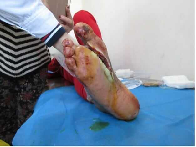
A 73-year-old man, with type 2 diabetes of 5 years’ duration, presented with an ulcer on the upper heel of the posterior tibia of the right lower limb right lower that was mostly covered with eschar that had been present for 6 months. He was admitted on March 18 for the management of surrounding erythema and swelling and administration of systematic antibiotics, and thereafter the wound was self-managed with traditional dressings.
The wound continued to deteriorate and become painful (Figure 2a), and he was referred on May 6 for further management. The wound measured 5.0 cm × 4.0 cm. On examination, the dorsal pedis and posterior tibial arteries of both lower feet were barely palpable. Initially, debridement was conducted (Figure 2b), the wound flushed with saline 0.9% and TLC-NOSF contact layer applied. The dressing was changed on alternate days. By June 25, the wound bed was looking healthier, and the wound had decreased in size (Figure 2c). The same management was continued by the family at home. The wound was almost completely healed by July 11 (Figure 2d) and completely healed in the following weeks.
Further management of the lower-limb ischaemia will be followed by the vascular team.
Case 3
A 65-year-old man, with a 26-year history of type 2 diabetes, sustained trauma on his right foot which went unnoticed initially due to peripheral neuropathy, with ensuing swelling and ulceration around the second toe. The patient self-managed at home with traditional methods. Within 2 months the wound worsened drastically, and the second toe became gangrenous. He was referred and the second toe of the right foot was amputated. He was discharged after his condition improved with systematic treatment. However, no further improvement was noted, and he was again referred with a deep right foot ulcer at the junction of the first and second metatarsophalangeal joints, with a large amount of purulent exudate and significant pain (Figure 3a). The wound was debrided and cleansed with 0.9% saline and TLC-NOSF contact layer was applied topically and changed on alternate days thereafter. Systematic antibiotics were initiated. By July 25, the wound was looking healthier, and the wound had decreased considerably in size (Figure 3b). The wound continued to progress. By August 10, the wound was progressing to healing (Figure 3c) and by August 29 the wound had closed (Figure 3d).
Conclusion
It is well know that diabetes-related limb complications pose a substantial clinical and economic burden to healthcare systems, as well as having devastating effects on patients and their families and carers (Lo et al, 2021). Overall rates of diabetes-related lower extremity complications have increased by 15.9% between 1990 and 2016, with the largest increases being recorded in southern sub-Saharan Africa, South Asia, and Southeast Asia (Zhang et al, 2020).
Regardless of the cause, it is essential that correct diagnosis and prompt evidence-based treatments are implemented promptly. Wound healing is a complex process, and even more so in people with diabetes. It is of utmost importance that holistic management is applied for these patients. The importance of local wound care using dressings with a strong evidence base has been given more importance in recent years. This has been highlighted in many publications, including recent studies, such as the Explorer trial, and recommendations from the IWGDF and the UK’s National Institute for Health and Care Excellence (NICE; Edmonds et al, 2018; NICE, 2019; Schaper et al, 2019).
The cases discussed represent a small cohort of patients with DFUs which were managed under real-life conditions in Harbin, China. The results obtained with TLC-NOSF characterise a fast improvement in the wound-healing process through wound surface area reduction and time to wound healing. The results of these three clinical cases are consistent with findings in other publications outlined in the systematic review by Nair et al (2021). The results suggest that TLC-NOSF provides a tangible clinical benefit for chronic wounds, including DFUs, and is an evidence-based, viable choice in their management in conjunction with an evidence-based standard of wound care.


