
The Masquelet technique is a surgical tool used in the treatment of bone defects in long bones. This technique was started in 1986 by Alain Masquelet and his team at the Avicenna hospital in France. They developed a technique for reconstructing bone defects. This technique is well known and well researched, used for the treatment of extensive bone loss due to severe trauma, tumours and infection (Romero-Berrío et al, 2021), and its procedure includes two steps.
The first is meticulous surgical debridement followed by the placement of a poly(methyl methacrylate)(PMMA) spacer implant, to cover the void by preserving neurovascular structures and healthy bone and soft tissues; temporary fixation of the spacer is done by internal or external fixation and after 4 to 8 weeks, the second procedure is performed by removing the cement spacer, performing repeated debridement while preserving the membrane and inserting an autologous bone graft with proper bone fixation (Luttenberger et al, 2020a).
There are currently only seven reports in the literature about bone defects of the foot treated with Masquelet technique, due to traumatic injuries of the heel cups and half feet. However, no publications or reports have been found about the use in patients with loss of metatarsal (short) bones due to osteomyelitis, as an infectious complication of the diabetic foot. The cases of short bones currently reported are in people without diabetes, which were around 10 case reports, and without osteomyelitis, generating a critical bone defect and micro-vascular inconveniences typical of the hand and foot. This type of treatment could validate the efficacy of the technique in the treatment of complications, specifically in osteomyelitis, which can be found in patients with diabetic foot through these case reports that involved open fractures, bite injuries and even gunshot injury. Optimal structural and functional recovery was consistently appreciated. These case reports have revealed that the Masquelet technique emerged as an alternative commonly chosen by patients who initially received an indication for amputation due to the complexity of their bone lesions, but that due to discomfort and limited or no acceptance by the patients, the “alternative” of amputation was chosen (Abdulazim et al, 2019; Andrzejowski et al, 2020; Jogani et al, 2020; Kasha and Yalamanchili, 2021; Luttenberger et al, 2020b; Qu et al, 2020; Saito et al, 2020; Liu et al, 2022; Encinas et al, 2023).
Diabetic foot ulcers are a frequent cause of morbidity, hospitalisation and amputations in patients with type 2 diabetes. These develop from the absence of neuropathic sensitivity or external trauma, such as exposure to repetitive periods of stress generated from walking, and they are aggravated due to ischaemia, neuropathic infection and oedema, which play a role in the development of ulcers that are difficult to heal, in addition to their pro-inflammatory environment (Moreno et al, 2013). Osteomyelitis is an infection frequently associated as a cause of a torpid evolution of neuropathic ulcers in patients with diabetes mellitus. This group of patients show a greater tendency to soft tissue and bone infections, compared to those without diabetes.
Infected ulcers significantly increase the risk of amputation (Orellano et al, 2022), this being the most frequent complication of diabetic foot (DF), and the main trigger for minor and major amputations. Osteomyelitis (OM) is present in up to 60% of cases and its treatment is a challenge, generating controversies depending on the different clinical presentations. Resection of the infected bone has been the standard treatment, and can generate functional sequelae and recurrent ulcers, complicating the prognosis of these patients (Romero-Berrío et al, 2021).
The purpose of this report is to propose the Masquelet technique as an alternative treatment of metatarsal osteomyelitis as a complication of diabetic foot. The results of this report could positively impact the prognosis and quality of life of these patients, especially in the prevention of amputation and in the postural improvement of the patient experiencing these complications (Encinas et al, 2023).
Case description
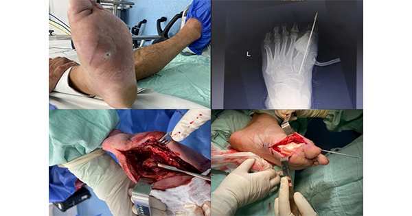
A 59-year-old patient entered the Emergency Department of Clínica de Occidente, Cali, Colombia, due to pain in his left foot, who upon inspection and palpation shows oedema associated with inflammatory changes at the medial and distal sides of the sole. He was ordered to undergo admission tests, including a foot X-ray, showing osteomyelitis of the head of the first metatarsal bone and the first phalanx. The results of the relevant laboratory tests showed a glycosylated haemoglobin (HbA1c) level of 8.2, with a plethysmography of 0.90 for right lower limb and 0.98 for left lower limb.
The San Elian Classification was used for the assessment and classification of diabetic foot, which gave a score of 10 [Table 1] (Moreno et al, 2013). A medial plantar ulcer treated on multiple occasions in different institutions, without favorable clinical evolution was found on the medical history. Based on the clinical history and paraclinical findings, the appropriate course was proceed with surgery to perform the necessary debridement, performing an osteotomy of the first metatarsal bone from its midsection to the head, and the first phalanx.
Due to the patient’s current conditions, it was decided to perform the Masquelet technique. Given the presence of osteomyelitis and the nature of the injury in the first metatarsal bone, this technique was chosen to give the patient an opportunity for a favourable prognosis without needing amputation [Figure 1].
The Masquelet technique was applied as follows:
The poly(methyl methacrylate) cement spacer is placed after the debridement and curettage procedure, and has two functions:
- A mechanical and structural support function to preserve the continuity and length of the skeletal segment, preventing tissue invagination. This can be done with mini and special external fixatives for short bones, such as those of metatarsal bones
- A biological function, based on the rich vascular content and its bioactive role. This membrane acts by promoting groups of vascular and ontogenic inducing factors to promote the formation of a suitable bone callus (Mejail and Caviglia, 2016).
- The reconstructive principle is based on promoting suitable local conditions to configure a perfectly delimited neo-compartment between the remaining bone stumps. This compartment must also be capable of being seeded a second time with bioactive material to promote osteogenesis (especially autologous bone, eventually increased with autologous bone marrow concentrate, bone substitutes or allogeneic grafts). Two specific requirements for this patient are highlighted:
- The compartment must have well-defined boundaries both circumferentially and at the implantation bases in both bone stumps
- The interior wall of the compartment must have fertile biological conditions for reception.
At approximately week three, the biological contribution has the highest levels of growth factors and adult stem cells within the membrane. At this stage, it is reasonable to infer that the optimal biological conditions for seeding the graft have been established. The spacer is exposed by longitudinal incision of the induced membrane.
The spacer is removed, preserving the membrane as a sleeve. As in the bed preparation, the use of instruments that promote tissue necrosis, such as thermocautery, should be avoided. The ideal biological contribution is the spongy autograft fragmented into a piece according to the extent of the defect, such as the iliac crests, or by the extraction of bone material from the proximal part of the tibia as it is a spongy and good quality bone. They can be extracted by the Reamer-Irrigator Aspirator System (RIA), but the latter depends on the case since if there is no other bone involvement, it is not necessary to do so. In this case, it was unnecessary because the bone is very small and does not require the use of a reamer. The only things used were the curettes and/or very thin bits. Masquelet explicitly clarifies that this proportion is an empirical notion (Mejail and Caviglia, 2016). The following are considered fundamental technical details: a) the block closure of the membrane and surrounding tissues; and b) the focal drainage since it is mandatory to avoid bruising, the space is required to be closed after surgery. This technique has been carried out where the smallest bone is a radius, the reconstruction of post-traumatic segmental bone defects using membrane induction technique (Klaue et al, 2009; Masquelet and Begue, 2010; Obert et al, 2016).
Autologous iliac crest graft is considered the gold standard, as a source for bone graft in Masquelet surgical technique, due to different factors. The main one is the relatively high concentration of mesenchymal cells (MSC), which can differentiate into other types of cellular tissues including osteoblasts, as well as their abundant amount of growth factors, such as TGF-B (transforming growth factor beta) and PDGF (platelet-derived factor), which play a role in the proliferation of bone cells (Masquelet et al, 2000; Pelissier et al, 2004).
Membrane induction surgical technique
Debridement — first step:
Devitalised bone fragments, sequestrations, devitalised or necrotic soft parts, fistulas and fibrosis are eliminated, verifying that there is no infection. Subsequently, the spacer is prepared. Bone cement was used, routinely applied in cemented arthroplasty, which has two components: 40g of polymer powder, consisting mainly of poly(methyl methacrylate) (PMMA), and 20 millilitres of liquid monomer, consisting mainly of methyl methacrylate (MMA). Four grams of one or more thermostable powder antibiotics (clindamycin, vancomycin, meropenem and ceftazidime) were incorporated into PMMA, chosen based on the following criteria: 1) culture result and antibiogram, 2) in chronic osteomyelitis with negative culture (vancomycin can be chosen to target S. aureus), 3) in acute bone loss due to exposed fracture without evidence of infection; vancomycin is an antibiotic commonly used in the Masquelet technique, due to its effective antimicrobial properties against Gram-positive bacteria, including methicillin-resistant S. aureus, a common agent in bone infections. On the other hand, it also has a great capacity to be consistently released from bone cement, which provides a localised concentration of the antibiotic at the site of infection helping to prevent bacterial growth. It is important to mention that experiments have also been carried out with other aminoglycoside antibiotics, such as gentamicin, with similar results.
Unfortunately, studies aimed at determining the particularities of antibiotics have thus far been conducted primarily in animals, imposing limitations on the robustness of the evidence they can provide. That said, these studies have suggested that the preference for vancomycin and gentamicin is supported by cellular and molecular foundations. It has been observed when gentamicin is used in concentrations greater than 100 µ/ml, it significantly impairs the number and activity of osteoblasts, but when the dose is lower, it acts almost as an additional osteoinductive factor. In contrast, vancomycin generally exhibited lower toxicity. The adverse effects found with gentamicin only manifested when its dose was increased up to tenfold (Giannoudis et al, 2011; Pelissier et al, 2002; Vejarano-Solano et al, 2015; Masquelet et al, 2019; Wang et al, 2019; Bilichtin et al, 2020; Meng et al, 2020).
Spacer removal
Between 4 and 8 weeks later (in this case, it could not be performed during the third week due to administrative barriers within the health system that prevent prolonged hospitalisation), the spacer can be removed, ensuring the induced membrane remains intact. Upon inspection, the induced membrane had to show the following characteristics: thickness 1-3mm; internal surface not adhered to the spacer, epithelial appearance and pale reddish colour, and external surface adhered to the upper plane tissue.
Graft contribution
Spongy bone graft is used taking the anterior iliac crest or tibia as the main source. Serial radiographic checks should be performed on patients, documenting the time in which consolidation was observed and the time in which full loading was allowed (Vejarano-Solano et al, 2015).
Definitive stabilisation
Definitive stabilisation must be carried out with external or internal fixation. Spongy bone graft is used, taking the anterior iliac crest or tibia as the main source. Serial radiographic checks shall be performed on patients, documenting the time in which consolidation was observed and the time in which full loading was allowed (Vejarano-Solano et al, 2015).
In orthopaedics, once the appropriate assessments are obtained, surgical treatment is done based on the findings from the examinations. Debridement is performed and the compromised bone part is removed (we do it in the first or fifth metatarsal bone). The cement part is placed on the methyl methacrylate with antibiotics and the area must be left closed so that the induced membrane is formed. Following this, and with the antibiotics prescribed by infectiology, negative pressure therapy is applied, and the process continues accordingly.
Difficulties and precautions of this technique in people with diabetes and osteomyelitis
The complications and precautions that should be considered with people with diabetes include the following:
- In cases involving this patients with infectious osteomyelitis affecting a metatarsal bone, the difficulty arises from their being very small spaces, which complicates their management and, therefore, the application of the technique (Obert et al, 2016)
- Not making a stable fixation. This means that the integration of the graft can be delayed in its consolidation or that it is not reached
- Skin coverage, if needed, is complex, due to the patient’s compromised micro and macro-vascular conditions (Obert et al, 2016)
- Careful consideration must be given to the selection of drugs suitable for these patients, due to their underlying clinical condition.
Following the implementation of the Masquelet technique as a treatment, the patient was discharged with a cast splint, a walker boot, and provided with all the medication required for his underlying pathology and comorbidities, as well as scheduled controls with enterostomal therapy (ET).
At 4 weeks the X-ray shows graft preservation and the patient is walking with support using his walker boot without showing any signs of an infectious process.
Another surgery was required after 7 months due to inadequate immobilisation [Figure 2]. During this surgery, which showed no signs of infection, spongy grafts were placed on the proximal tibia, and a plate was used to achieve better fixation. Also, a plaster splint and walker boot.
The patient is currently walking on his own, without any inflammatory change at an external level, and is in the process of graft consolidation. A third procedure was necessary because the patient failed to use the prescribed restraints.
Discussion
Current literature does describe the use of the Masquelet technique, however, in patients diagnosed with diabetic foot it has only been applied in long bone lesions (femur and tibia). There is no experience in the application of this technique in patients with osteomyelitis, or with the aim of improving the quality of support and functioning. Only one case has been reported in the Revista de la Sociedad de Ortopedia y Traumatología (SCCOT), but this case is of traumatic origin.
The patient in this case report had undergone two to three surgical procedures in other institutions, but these treatments only focused on the release of purulent material. When he presented at the Clínica de Occidente de Cali, foot X-rays of his foot were taken, as indicated by our protocol. Based on these X-rays he was diagnosed with osteomyelitis, so it was decided to perform exhaustive debridement of the lesion, drying out the distal half of the first metatarsal bone and the proximal phalanx of the hallux. Based on the results of the initial procedure, the possibility of employing this surgical technique was considered, which was aimed to give the patient an opportunity to avoid major complications and prevent amputation. Among the potential limitations that could hinder the application of this technique were administrative constraints, influenced by the dynamics of the Colombian Health System, such as the timing of cement removal with antibiotics, which is removed after 6 days. However, as described, this did not impact the outcome.
The outcome for the patient was a satisfactory, but unfortunately required new surgery 7 months after the procedure because the patient did not adequately use the restraint kit for a long time. Given this, new surgery was required to place spongy grafts of the proximal tibia and a plate to make better fixation, with a plaster splint and walker boot.
As specified above, timing issues within the health system are crucial, and they can, to a certain extent, hinder the achievement of more beneficial results for patients. To solve this issue, it has been proposed to extend the duration of cement with antibiotics to achieve a more appropriate induced membrane by discharging the patient for a period of 4 to 6 weeks, and returning to the second procedure, maintaining outpatient controls. In this way follow-up could be carried out combining the areas of service provision.
Subsequently, during the follow-up controls, a control X-ray was conducted at 4 weeks, showing preservation of the graft, the patient is walking with support using his walker boot without signs of an infectious process.
Due to the results obtained with this patient and with the conviction that this type of procedure can benefit multiple patients with this type of complications, such as diabetic foot ulcer, we believe that more studies with greater methodological validity would be necessary to generate more knowledge of the applicability of this technique in these patients, generating more specificity in the indications for these pathologies, knowledge and new therapeutic alternatives in the specialty of orthopaedics.
Conclusions
The Masquelet technique is a technique that we should implement in diabetic patients, as it can improve their quality of life, by effectively managing the complications associated with osteomyelitis, it helps preserve the patient’s functionality.
As the Masquelet technique was initially implemented for large bone defects, evidence on its implementation in short bones is limited. However, during the literature review for this article, 10 case reports were found documenting its efficacy in short bones, confirming its efficacy and replicability in different patients, hospital contexts and aetiologies of the critical bone defect that allowed the indication of the Masquelet technique.
To apply this surgical technique in patients with diabetic foot with osteomyelitis, they must have skin without necrosis, which is why an adequate environment must be guaranteed to be able to completely close the intervened part.
They must be immobilised properly and safely to allow the formation of a robust bone callus. Orthopedists know that any fracture requires 0.05 microns of mobility for consolidation, and if this is exceeded the fracture may develop into pseudoarthrosis.
It is essential for patients with diabetic foot to maintain proper metabolic control of their underlying disease and the orthopaedist should consider monitoring total proteins, albumin, globulins, as well as levels of vitamins B12 and D3 in these patients, along with other paraclinical test necessary to ensure an optimal evolution.
In terms of medication used, these were pregabalin and 600mg alpha lipoic acid (an excellent hydro- and fat-soluble antioxidant) about 45 minutes before or after breakfast and their respective assessments, to improve oxidative stress which, in turn, helps manage inflammation.
Periodic outpatient controls were initiated to carry out adequate follow-up, so that the patient adheres to their prescribed treatments.
Despite the lack of evidence and reports of experiences with the Masquelet technique in the reconstruction of bone defects in the foot, it has been demonstrated in this case that there are encouraging outcomes for the use of this surgical alternative, considering the important relationship between the restitution of foot structures and the restitution of patient functioning.


