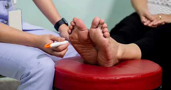
Plantar foot ulcerations are a common source of morbidity in patients with diabetes and peripheral neuropathy. An estimated 15% of patients with diabetes will suffer from lower extremity ulceration during their disease process (Frykberg et al, 2006). Approximately one-third of all diabetic foot ulcerations (DFUs) occur on the plantar surface of the hallux (Ctercteko et al, 1981; Armstrong et al, 1998; Peters et al, 2007). This and the plantar first metatarsal head are the most common locations for neuropathic ulcers to develop. In a retrospective study of 360 patients treated for diabetic foot ulcers, Armstrong et al (1998) found that 30% of these ulcers occurred on the plantar hallux. A similar study by Peters et al (2007) found 24 out of 81 (30%) of diabetic ulcers to occur under the first ray, 18 of which were located on the plantar hallux. Ctercteko et al (1981) also demonstrated that the majority of plantar ulcers occur on the medial aspects of the hallux and first metatarsal head, in the area of maximal loading.
Not only are hallux ulcerations common, they are also difficult to heal and frequently recur. The prospective cohort study by Peters et al (2007) evaluated the risk factors contributing to re-ulceration and found peripheral arterial disease (PAD) and ulcer location to be significant. Of the 81 patients in this study, 18 suffered a plantar hallux ulcer which was significantly more likely to re-ulcerate.
Frykberg et al (2006) discussed multiple risk factors contributing to diabetic foot ulcerations including peripheral neuropathy, vascular disease, limited joint motion, foot deformity, abnormal plantar pressures, minor trauma, a history of ulcer or amputation, and poor visual acuity.
In a prospective analysis of 33 feet with plantar hallux ulceration, Boffeli et al (2002) found that nearly all had severe ankle equinus, marked hallux limitus, and hallux interphalangeal abductus. In a similar study, Nubé et al (2006) found the plantar hallux ulceration to be associated with a more pronated foot type. In addition, when comparing patient’s feet with hallux ulceration, there was no difference in the amount of first metatarsophalangeal (MTP) joint dorsiflexion. On the other hand, when comparing first metatarsophalangeal joint dorsiflexion in the affected and unaffected foot of the same patient, the study found approximately a 10° reduction in motion in the ulcerated foot.
Conservative care using an orthotic device can reduce the pressure under the plantar surface of the hallux (Wu et al, 2005). While effective in offloading the great toe when used, the associated hallux limitus/rigidus still obviously remains. Those with plantar neuropathic ulcerations may also be treated with other offloading modalities including total contact casting, fixed ankle walkers, healing sandals, half or wedge sole shoes, felted foam, and multidensity inserts (Wu et al, 2005).
A review by Frykberg et al (2010) of surgical offloading procedures for diabetic foot disorders discussed the value of the first MTP joint arthroplasty (Keller arthroplasty) for the management of wounds located at the plantar aspect of the hallux interphalangeal joint. In the setting of chronic open ulcerations, such procedures have been termed as “curative”, since they are performed primarily to effect healing of otherwise recalcitrant wounds (Frykberg et al, 1993; Armstrong and Frykberg, 2003; Armstrong et al, 2006).
In 1904, William Keller was the first to describe his bunionectomy involving the resection of the base of the hallux proximal phalanx (Keller, 1904). The Keller arthroplasty has traditionally been indicated for painful hallux valgus and hallux rigidus with associated degenerative joint disease in the older population (Keller, 1904; Rankin and Rankin, 1996; Stewart and Reed, 2003).
There are several benefits to this procedure including pain relief, correction of deformity, resolution of skin lesions, and permitting a quick return to activity postoperatively. It is generally accepted that increased first metatarsophalangeal (MTP) joint dorsiflexion achieved with the Keller arthroplasty can result in healing of plantar hallux ulcers with minimal complications. Nonetheless, every procedure has its potential complications. These include transfer metatarsalgia or ulcerations, short toe, loss of toe purchase, floppy or cocked up hallux and weakness during the push-off phase of gait (Keller, 1904; Downs and Jacobs, 1982; Stewart and Reed, 2003; Berner et al, 2005; Beertema et al, 2006).
Despite these complications, the Keller arthroplasty has been demonstrated as a reliable procedure for correcting hallux valgus deformities with underlying degenerative joint disease, as well as for those persons with hallux limitus or rigidus disorders (Rankin and Rankin, 1996; Beertema et al, 2006). Furthermore, experience has shown that this procedure is a reasonable alternative to amputation of the hallux or first ray in patients with chronic plantar hallux ulcerations (Armstrong et al, 2003; Berner et al, 2005).
This retrospective study aims to review the authors’ experience with the Keller arthroplasty in both ulcerated and non-ulcerated patients for the treatment of hallux valgus or hallux limitus/rigidus conditions. The authors will contrast healing times as well as post-surgical complications between diabetic and non-diabetic patients as well as between those with and without peripheral neuropathy. Full approval of the Phoenix VA Institutional Review Board was obtained prior to initiating this study.
Patients and methods
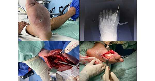
A retrospective medical record review of 54 consecutive patients (57 feet) who underwent a Keller arthroplasty for a chronic plantar hallux interphalangeal joint ulceration (Figure 1) or primarily for clinical hallux valgus or limitus/rigidus conditions was performed. Any patient with suspected or confirmed osteomyelitis of the great toe or first MTP joint was precluded from having this operation. By virtue of its retrospective nature, we did not routinely perform range of motion or pressure measurements on patients preoperatively or postoperatively. All surgeries were performed by the senior author (RGF) at the authors’ institution between 2004 and 2012 with a mean follow-up of 37.6 ± 21.5 months.
The authors’ surgical technique was fairly standard in that dorsal midline incision over the first metatarsal neck and head that is carried distally to the mid-shaft of the hallux proximal phalanx was employed. After proper exposure, an inverted “L” capsulotomy is performed where the vertical arm of the capsular incision is carried along the medial phalanx distal to the base. The capsule is reflected off the phalangeal base and metatarsal head to allow resection of the base, medial metatarsal eminence, and any other exostoses present on the metatarsal head. The sesamoids are mobilised through a combination of sharp and blunt release of capsular adhesions. Most frequently, a straight transverse resection of the phalangeal base is performed approximately 1 cm distal to the joint.
Care is taken not to resect too much bone nor too little bone to avoid excessive laxity or subsequent restriction of motion, respectively (Figures 2 and 3). For severe hallux valgus we will make an oblique cut, leaving a longer lateral portion that can articulate with the laterally deviated cartilage position on the metatarsal head. For capsular repair, we attach the tissue to the remaining phalanx through two medial/dorsal drill holes while holding the toe in corrected position with non-absorbable sutures. We rarely require a K-wire to stabilise or correct the toe position, except in the case of severe deformity. On the contrary, we do not use internal hardware in the presence of an open ulceration.
Care was taken to exclude osteomyelitis, probing to bone, or active infection of ulcers at the time of surgery. Accordingly, superficial ulcers were covered with an adhesive polyurethrane barrier during the procedure. After closure of the incision and application of a barrier dressing, the ulcers were debrided, cultured, and dressed separately from the incision. The authors routinely administered a single prophylactic dose of cefazolin or clindamycin preoperatively; this was followed by oral therapy only when ulcers were present. Postoperatively, patients were fitted with a standard postoperative shoe and were ambulatory upon release from their outpatient surgery.
The average age of the patients was 67 ± 7.6 years old, 98% of whom were men. Thirty-two (59.3%) of the patients had a diagnosis of diabetes, but only 4 (7.02%) had PAD based on history, absence of pulses, or abnormal non-invasive studies preoperatively. At baseline, 28 (51.8%) patients were found to have peripheral neuropathy, defined as having loss of protective sensation to the 10 g monofilament, insensitivity to pain, and/or lack of vibratory perception at the forefoot level. Current smokers comprised 15% of the study population, while nearly 60% of patients were previous smokers. Only 25% of subjects had never smoked. Twenty-eight of the 57 feet (49.1%) had a diagnosis of hallux limitus (restriction of dorsiflexion with or without degenerative changes of the first MTP joint), 13 hallux rigidus (22.8%), 15 hallux valgus (26.3%), and one with combined hallux valgus and hallux limitus (1.8%). Fifteen (26.3%) patients had an ulcer present at the time of surgery, and 13 of these 15 patients (86.7%) had diabetes. Patient demographics are listed in Table 1.
Chi squared, two sample t-test, and logistic regression analyses were used as appropriate to investigate predictive factors for healing and complications. Patients were queried at examination or by telephone interview concerning their satisfaction with the outcome of their procedures. All analyses are based on individual feet undergoing this operation unless otherwise noted. The outcome was considered statistically significant if P<0.05.
Results
Of the 57 procedures performed, 45 (79%) healed uneventfully, while 12 patients (21.0%) experienced complications postoperatively. Those complications observed were primarily infection or wound dehiscence. None of the patients required an amputation prior to healing, however. Although there were more complications in patients with diabetes, the frequency did not reach statistical significance (27.3% versus 12.5%, P=0.177). However, neuropathy was significantly associated with the development of postoperative complications (34.5% versus 7.14%, P=0.01), as shown in Table 2. Accordingly, there was a seven-fold risk for post-surgical complications in those patients with peripheral neuropathy (OR 6.8, 95% CI 1.34–34.89).
Sixty-seven percent (8/12) of complications occurred in feet with an open hallux ulcer (P=0.000). The authors found that the presence of an ulceration at the time of surgery imparted nearly an 11-fold risk for postoperative infection/dehiscence (OR 10.8, 95% CI 2.55–46.09; Table 3). Since 93% of the cohort had palpable pedal pulses, a significant association with PAD could not be demonstrated (P=0.176). While neither current smoking (P=0.19), nor age (P=0.84) were associated with the development of a postoperative complication in these patients, 8 of 12 (66%) patients who developed complications were prior smokers. On multivariate logistic regression where peripheral neuropathy was controlled by ulceration, only the presence of a hallux ulceration at the time of surgery predicted a seven-fold risk for the development of a postoperative complication (OR 6.86, 95% CI 1.12–41.83; Table 4).
While not statistically significant, people with diabetes also took a longer time to heal the incision site compared to those without diabetes (35.6 days versus 29.4 days, P=0.496). As would be expected, there were significantly more ulcerations present in patients with diabetes (13 versus 2, P=0.009). Two of the 15 patients with hallux ulcers were not diabetic, but had neuropathy secondary to chronic alcohol consumption.
In fact, all patients with preoperative hallux ulcerations had documented peripheral neuropathy at the time of surgery. While the time for hallux ulcerations to heal averaged 22.2 days, surgical incisions in these patients took more than twice as long to heal (50.2 ± 57.54 days) due to the development of incisional dehiscence or infection. People without ulceration healed in a mean time of 28.7 ± 18.7 days, significantly shorter than those with ulcers (P=0.05).
Average shortening of the hallux following Keller arthroplasty for the entire cohort was 7.3 ± 3.5 mm as measured on digital radiographs. Transfer lesions were infrequent, although the authors often noted hammering of the second toe as a consequence of the procedure. The median time to a return to customary footwear was 32 days (interquartile range 12–91 days) with a mean of 43.2 ± 35.8 days. However, in this elderly population, a 94% overall self-reported satisfaction with the procedure was reported.
Discussion
Hallux rigidus and hallux valgus are common foot conditions that require surgical intervention for relief if they are not responsive to conservative treatment. Arthrodesis has been defined as the gold standard for hallux rigidus and a proximal metatarsal procedure has been defined as the gold standard for severe hallux valgus. Due to the elderly patient population and those with multiple co-morbidities at the authors’ institutions, the authors have sought other surgical treatment options for these conditions. Additionally, many of the authors’ patients, due to physical impediments, cannot be relied upon to be compliant with the rigorous post-operative care of an arthrodesis of the first MTP joint or first metatarsal-cuneiform joint.
The Keller arthroplasty has been advocated as a procedure for hallux valgus and rigidus in the elderly population with degenerative changes within the first metatarsophalangeal joint. The authors find that the procedure indeed proves to be a viable option for degenerative hallux valgus, hallux limitus/rigidus, as well as for the recalcitrant plantar hallux ulceration.
One criticism of this arthroplasty is that it is cosmetically displeasing, often having an end result of a short, fat, floppy toe. Modifications have been proposed to limit this problem including surgical techniques to interpose capsular components and utilising different fixation methods to maintain length (Thomas, 1962; Mroczek and Miller, 2003; Kolker and Weinfeld, 2007). The authors’ surgical technique for maintaining length and obtaining a more cosmetically pleasing result is either to interpose capsule via the purse-string technique or attaching the dorsal capsule to the remaining base of the proximal phalanx of the hallux. By doing one of these two techniques, patients at the authors’ facility appeared to be satisfied with the postoperative result. Average shortening of the hallux in this study was only 7 mm. This shortening seems to be more of a cosmetic concern and not a functional one. McLaughlin and Fish (1990) reported in their study the use of distraction with a K-wire following the arthroplasty and found an overall improvement of symptoms, function and cosmesis not dependent on the type of procedure utilised. Hahn et al (2009) found no statistical significance between physical function pre-operatively and postoperatively following this procedure with an interpositional capsular repair.
Functional hallux limitus, as described by Dananberg (1986), refers to a condition of the first MTP joint, wherein there is a restriction of dorsiflexion upon weight-bearing in the absence of radiographic signs of degenerative joint disease. Quite frequently, this restriction of hallux motion during stance and gait is an underlying cause for high plantar pressures leading to ulceration. Operations, such as the first MTP arthroplasty, are aimed at increasing the available range of hallux dorsiflexion and thereby allow for healing of otherwise recalcitrant great toe ulcerations (Armstrong et al, 2003; Frykberg et al, 2010). This current study also confirmed that this procedure was associated with a fairly rapid progression to healing of these chronic hallux wounds.
A modified Keller arthroplasty has previously been advocated for “resistant” plantar hallux ulcerations (Berner et al, 2005; Beertema et al, 2006). Downs and Jacobs (1982) noted complete healing of plantar hallux ulceration within 33–35 days postoperatively in the six patients treated with this procedure. The only complication was delayed healing of the incision site. All wounds remained healed at 2–5 years of follow up.
A retrospective chart review by Berner et al (2005) of 13 feet in 11 patients demonstrated that complete healing of plantar hallux ulcerations was achieved with a modified Keller arthroplasty. Even though all wounds healed within 6 months and remained healed at 1 year, five of 13 feet in the study developed transfer ulcers.
In a 2003 case-control study of 41 patients with neuropathic ulceration under the interphalangeal joint of the great toe, Armstrong et al (2003) found that those patients treated surgically with a first MTP joint arthroplasty healed significantly faster (24 versus 67 days, P=0.0001) and had fewer recurrences than those treated with standard nonsurgical management. Interestingly, approximately 40% of their study population developed infection prior to healing.
The present study included plantar hallux ulcerations in 15 patients. As might be anticipated, there was a higher occurrence of hallux ulcerations in those patients with diabetes and neuropathy. Similar to the aforementioned study, these ulcerations healed in an average of 22 days following surgery. In contrast, Berner et al (2005) reported an average healing rate of 6 months for plantar hallux ulcerations following Keller arthroplasty. While all of the patients in their study had adequate vascular supply, 7% of patients had PAD. Regardless, the presence of PAD in this study was not found to be a predictor of wound healing delay nor postoperative complications. Lin et al (1996) retrospectively reviewed the results of Keller arthroplasty with total contact casting (TCC) and found no postoperative complications in an average follow up of 26 weeks, with a mean hallux ulcer healing time of 24 days. To the contrary, the authors’ present study found a higher rate of complications in patients with neuropathy or hallux ulcers, although the authors had a similar ulcer healing time of 22 days. Importantly, none of the complications led to an amputation during the follow-up period.
Limitations of the current study include its retrospective design, small sample size, focused/specific population studied (all but one were men older than 55 years undergoing the same operation) and non-uniformity of data contained in electronic medical records. Furthermore, the authors had no comparison group of patients having had alternative operative procedures.
Due to the small sample size, where no significant associations were detected in analyses, a lack of power to detect such relationships may have existed. This is especially true for the lack of association with PAD, a well-known risk factor for wound healing failure and infection. It is certainly probable that more patients presented with clinically demonstrable vascular disease, the authors would have found such an association to be significant. Nonetheless, more than half of the patients in the current study had diabetes, 73% of whom healed uneventfully.
Furthermore, the authors found that those patients at highest risk for complications postoperatively were those with neuropathy or ulceration. In those patients where such a curative operation is deemed necessary to heal a recalcitrant hallux ulcer, the authors find that the Keller arthroplasty is highly successful. In those patients with degenerative hallux valgus, rigidus, or functional hallux limitus in the absence of an open ulcer, this operation can also be performed quite successfully with minimum complications. To confirm the findings and to address the aforementioned weaknesses of this retrospective study, the authors are currently engaged in a prospective evaluation of this same operation in a larger group of patients.
Conclusion
In this retrospective study of the first MTP arthroplasty, a high degree of success with minimal postoperative complications was found. Most complications can be anticipated in those patients with an open hallux ulcer or neuropathy. As a whole, patients with diabetes were at no greater risk for infectious complications unless they had an ulcer present at the time of operation. Nonetheless, this operation led to fairly rapid healing of all existing hallux ulcerations without the need for amputation in any case. Despite the consequent shortening of the great toe, 94% of patients expressed satisfaction with the results of this procedure. Accordingly, the authors continue to favour the Keller arthroplasty as an effective intervention for patients with significant degenerative joint disease associated with hallux valgus or rigidus, or in those patients with plantar hallux ulcerations associated with functional hallux limitus.


