
Most amputations (85%) start with a single foot ulcer (Larsson, 1994). There is a high prevalence of digital deformities in diabetes patients, which can lead to the ulceration on the apex of the toe (Calvo-Wright et al, 2023). Toe deformities are common in diabetes and a common site of ulceration that can lead to digital or higher amputation. Toe deformities are caused by an imbalance between the extrinsic and intrinsic foot muscles (Boulton, 2025). It is vital that these are managed with the most suitable treatments, such as adequate footwear, silicones, callus debridement and ulcer management (Scire et al, 2009). Ulceration of the toes account for 43–55.5% of all diabetes foot ulcer cases, and while these ulcers are smaller and typically heal faster other location on the foot, they are often underestimated and tend to have higher rates of limb amputations compared to other foot locations (Pickwell et al, 2013).
The Internal Working Group on the Diabetic Foot (IWGDF) guidelines state that: “In a person with diabetes and a neuropathic plantar or apex digital ulcer, consider using digital flexor tenotomy to promote healing of the ulcer, if non-surgical offloading treatment fails” (Bus et al, 2023).
Percutaneous tendon release (tenotomy) of the flexor digitorum longus (FDL)/needle flexor tenotomies is a relatively safe and effective treatment for ulceration and to prevent formation of new ulcers associated with hammer, mallet and claw toe deformities (Anderson et al, 2019). There is a 95% healing rate after needle flexor tenotomies (Tamir et al 2014).
Clawed toes are defined as an extension of the metatarsophalangeal joint (MTPJ) and flexion of the proximal interphalangeal joints (PIPJ) and distal interphalangeal joints (DIPJ; Ledoux et al, 2008).
Pemayun et al (2008) outlines that 10–15% of diabetes foot ulcers will not heal and will need further intervention, such as amputation.
Case study
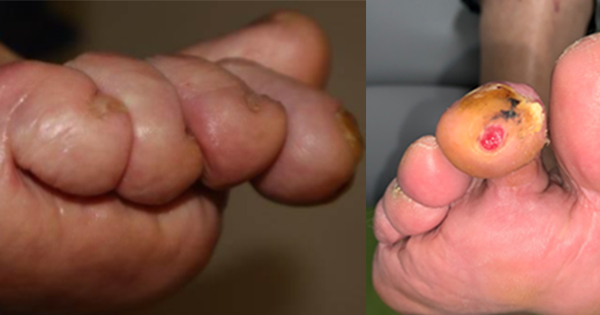
This case features a 67-year-old man, with type 2 diabetes since 2001. He is a retired engineer, married, two children, non-smoker and a social drinker with no reported allergies. He was referred to the diabetes multidisciplinary foot team (MDFT) in November 2021, as per the local diabetes foot pathway, with a chronic, non-healing apical ulcer on his right second toe since 2017. The aetiology was a combination of neurological changes within his foot with retraction and clawing of his right second toe, leading to ulceration. This had been treated for 12 months with footwear advice and a silicone toe support, but without full resolution.
Assessment was undertaken at the University Hospital Southampton MDFT. The MDFT consists of individuals from both community and acute hospital teams – podiatry, vascular surgeon, diabetologist, surgical care practitioner, microbiology and orthotist. The MDFT clinic is held once a week for acute foot disease due to diabetes. Access is via the commissioned diabetes foot pathway. The role of the MDFT in reducing amputation is well known (Krishnan et al, 2008).
On presentation, his HbA1c was 48 mmol/mol; a significant improvement from 9 years earlier, when it was 79 mmol/mol. His BMI was 32, with interventional advice given and referral to a dietitian. His blood pressure was 139/78 mmHg, total cholesterol 2.8 mmol/mol and estimated glomerular filtration rate 80. His medications were dulaglutide once weekly, and daily atorvastatin 20 mg, amlodipine 10 mg and ramipril 10 mg.
Assessment with the 128 Hz turning fork and 10 g monofilament showed absent vibration and monofilament from knee downwards on both limbs.
His skin was anhidrotic and showed structure changes, indicating motor, autonomic and sensory neuropathy.
The “prayers sign” is a clinical finding in neuropathy, and is assessed by asking the patient to put his or her hands together in a praying position with the fingers fanned and to press together the palmar surfaces of the interphalangeal joints and the palms (Upreti et al, 2003). This patient demonstrated a moderate prayers sign, which is involvement of three or more interphalangeal joints or one finger or one finger and one large joint bilaterally, and limited joint mobility (LJM). This is a condition characterised by glycation (Singh et al, 2014). The hand stiffness, resulting from flexion contractures of the fingers and thickened, tight, skin and is seen in approximately 25–50% patients with diabetes (Rosenbloom, 1989). The prayers signs is equally common in both sexes and in longer duration of diabetes (Ballantyne and Hooper, 2004).
On assessment, both feet popliteal, dorsal pedis and posterior tibia arteries were all palpable. With Doppler, all pulses strong biphasic. In addition, on Doppler, both peroneal and right medial plantar arteries were located. A toe systolic pressure and toe brachial index measurement, as recommended by Hinchliffe et al (2020), is routinely taken. His toe brachial index of 80 mmGH demonstrates no significant vascular issues.
A review of his footwear showed a well-worn lace up style shoe (Figure 1). He reported his footwear was not a problem as “they felt fine all of the time”.
On questioning, he reported never undoing the laces and “slipping them on”. This has the potential to cause pressure damage and could lead to his toes clawing more to keep the footwear in place. Footwear advice was given and instructions on to undo and retie his laces every time he puts them on.
Types of footwear was discussed, and a trainer style shoe was recommended. Cavanagh (2007) suggested the use of trainers or sports shoes as an alternative to custom-built footwear where these are not accessible or required.
Frykberg et al (2006) stresses the need for a musculoskeletal assessment that should include evaluation for any gross deformity. Observing the patient rising form the chair in the waiting room when called, he was able to slowly stand unaided. He took several seconds to get his balance on standing, which is an aspect of performance-oriented mobility assessment (Hong et al, 2017). On walking, his gait was slow, with a wide base of gait and prolonged double support time, consistent with observation from Wrobel and Najafi (2010) in their paper.
When he was observed standing barefoot, he had a wide base of gait and the right second toe made contact with ground, despite a silicone toe support being in use. He had a high arch foot, with loss of bulk of his intrinsic muscles on both feet.
He was asked to close his eyes while standing, and quickly became unstable with a sideways swing. This indicates his lack of proprioception due to his underlying diabetic peripheral neuropathy, which is attributed to loss of nociceptive feedback from the lower extremities due to small fibre degeneration (Malik, 2014). In addition, his toes clawed more when this was undertaken.
There was a restriction of the full 10% dorsiflexion at the ankle joint in both feet on both passive and active movement, with only approximately 5° of movement. This was consistent with tightness in the Achilles tendon. Glycosylation of soft tissues is the main caused of this, confirmed with his prayers sign (Wrobel and Najafi, 2010). There were clinical signs of diffuse callus over his first and fifth metatarsal heads indicating an increase in overloading of these sites.
Examining both feet in subtalar joint neutral, and loading the second metatarsal head, the right second toe was clawed more than the other lesser toes. On plantar flexion of the ankle, straightening of the toe was achieved, indicating soft tissue contracture and no fixed deformity at PIPJ and DIPJ.
His history showed that this ulcer had resulted in him being on several courses of flucloxacillin for mild infection and co-amoxiclav for moderate infection, as the ulcer previously been probing to bone, as per Trust and national guidance (NICE, 2019).
On presentation at MDFT, he had no signs of infection and the ulcer was classified as TEXAS A1 with a SINBAD score of 2/6, a moderate ulcer, indicating a healing time up to 77 days (Ince et al, 2008).
The silicone digital device can be clearly visualised on X ray (Figure 3). The patient had been using this for several months; however, this was found to be not effective in reducing pressure. Silicone padding is effective and safe in the prevention of lesions in neuropathic patients at high risk of ulceration (Scirè, 2009). So, once healed, silicones are effective.
The diagnosis was apical neuropathic ulceration due to clawing of right second toe, due to repeated trauma and pressure when the foot is in contact with the ground.
Intervention at MDFT
The right second toe was able to be straightened, indicating suitability for a FDL tendon release to extend the toe, remove the cause of the pressure from the ulcer site and resolve the ulceration. His present ulceration puts the toe at risk of infection and amputation. This procedure would be curative (Frykberg et al, 2020). The procedure was explained to the patient.
The percutaneous tendon release of the FDL was performed in clinic without the need for a local anaesthetic with a white needle (15 G). Incision was on the plantar aspect of the PIPJ. The tendon was successful divided after insertion by moving the white needle medially and lateral to tear the FDL under extension of the toe. Mechanically, this results in the toe becoming extended at the PIPJ with the apex no longer in contact with the ground forces and achieving full offloading of pressure on the ulcer site.
At the 6-week follow-up, the extension of the right second toe had achieved the full reduction in apical pressure and allowed the full resolution of the ulcer (Figure 4). The result was a straight and dorsally displaced toe, that can be seen both from the X+ray and external examination. There is ongoing monitoring and the patient has changed to wearing trainers.
The patient remains healed and ulcer free 3 years post his FDL tendon release.
Discussion
The combination of the position of this patient’s toe and his sensory neuropathy resulted in him having a high risk of foot ulceration (NICE, 2017). Pressure management of this toe was key because focus of peak pressures can be in excess of 1,000 kPa and that the repeated pressure insult combined with the force of shear, leads to tissue breakdown and foot ulceration (Russell, 2015). Due to the continual pressure, this resulted initially in callus formation which then progressed to ulceration. The duration of this ulceration poses a significant risk of amputation of his toe. Brownrigg et al (2013) highlighted that a foot ulcer is the initial event in >85% of major amputations that are performed on people with diabetes, so any intervention that prevents or heals a foot ulcer will be preventing that amputation.
His treatment prior to the MDFT assessment consisted of regular debridement to reduce the callus and pressure, which Edwards and Stapley (2010) indicate as a key treatment, alongside management of infection (Lipsky et al, 2012) and the use of a silicone removable offloading device (Bus et al, 2023). Numerous studies and international guidelines state that to achieve healing from a foot or toe, appropriate support and deflection of pressure will be required (Bus et al, 2020).
Fundamentally, peripheral neuropathy predisposes the foot to ulceration through its effects on the sensory, motor and autonomic nerves (Boulton et al, 2008). Clawed toes are associated with the foot in people with diabetes (Donatelli, 1995). The duration of excessive stress on tissue is one of the of the leading causes of diabetic foot ulcers among people with diabetic peripheral neuropathy. Lazzarini et al (2019) describe the concept of plantar tissue stress (PTS) to “integrate several well-known mechanical factors into one measure, including plantar pressure, shear stress, daily weight-bearing activity, and time spent in prescribed offloading interventions (adherence)”. The overall aim is to redistribute plantar pressures to prevent callus formation. Silicone props may be used to divert pressure (Bus et al, 2019).
In our case, repetitive mechanical forces of gait and the clawed position of his toe had led to the callus formation. As the callus forms and thickens, the callus will increase the tissue stress press on the soft tissues underneath and cause ulceration. If the callus is not removed, inflammatory autolysis and haematomas develop under the callus. This leads to tissue necrosis, resulting in a small cavity filled with serous fluid giving the appearance of a blister under the callus (Edmonds, 2002).
The IWGDF guidelines outline that offloading is the most important aspect when preventing and managing ulceration in a patient with diabetes (Bus et al, 2023). Cavanagh et al (2005) stated that alleviation of the mechanical load on ulcers (offloading) should always be a part of treatment.
Claw toe deformities are produced by a muscular imbalance between the extrinsic and intrinsic foot muscles (Boulton, 1988). It is vital that these are managed with the most suitable treatments, such as adequate footwear, silicones, callus debridement and ulcer management.
A small pilot study by Ledoux et al (2008) demonstrated that people with diabetes with neuropathic feet with claw toes have less intrinsic muscle volume than the other groups. Bus et al (2002) examined both sensory and motor neuropathy, and found that motor neuropathy in the feet to be profound in people with diabetes and stated that intrinsic muscle atrophy does not necessarily appear to cause toe deformity.
Tamir et al (2014) showed a 95% healing rate post needle flexor tenotomies. The use of digital flexor tenotomy is included in the most recent IGWDF guidelines (Bus et al, 2023).
Conclusion
In this case, needle flexor tenotomy was successfully undertaken. These are relatively safe and effective treatment compared to tenotomies done by scalpel, both as treatment for ulcers and to prevent formation of new ulcers associated with claw toe deformities.
Due to his significant neuropathy and glycation of his tissue, this patient will be always be at high risk of foot ulceration. Footwear and insoles will need to be provided for prevention of ulceration. Regular assessment to ensure that the glycation of his Achilles tendon does not lead to further plantar ulceration of further apical ulceration as his toes continue to claw.
MDFT assessment of all diabetes-related foot complications is key to the management and onward healing and resolution as clearly defined in NICE (2017) guidelines.
This case study identified a neuropathic chronic apical ulceration caused by pressure due to the position the toe had adopted. It is critical that the MDFT takes into account the biomechanical aspect of the foot in their assessment and treatment options.
This straightforward outpatient procedure undertaken within the MDFT setting led to resolution of the ulceration by removing the cause, a claw toe.
FDL tendon release is clinically indicated in apical ulceration in toes that can be extended with no fixed deformity in the MPJ, PIPJ and/or DIPJ, and where non-surgical offloading treatment has failed.


