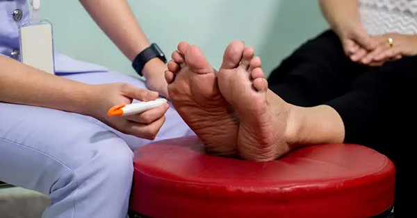
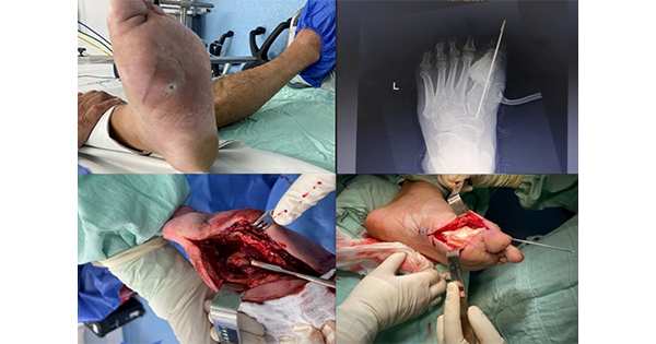
The protracted healing often seen in diabetic foot ulcers (DFUs) may be due to a multiplicity of often inter-related contributory factors, including excessive exudate production. Excessive exudate production by any type of wound may cause problems that ultimately delay healing, reduce quality of life for patients and place additional burdens on healthcare systems. Effective management of exudate may aid healing, reduce the impact on the patient of problems such as leakage and periwound maceration, and make more efficient use of healthcare resources.
What is exudate?
Wound exudate has had numerous descriptions, including ‘what is coming out of the wound’, ‘wound fluid’, ‘wound drainage’ and ‘an excess of normal fluid’ (World Union of Wound Healing Societies [WUWHS], 2007). It is derived from the fluid that leaks from blood vessels in the wound bed and first appears during the inflammatory phase in response to tissue injury as part of the normal wound healing process.
Role of exudate in wound healing
Wound exudate contains a variety of substances, including water, electrolytes, nutrients, inflammatory mediators, white cells, protein-digesting enzymes (e.g. matrix metalloproteinases [MMPs]), growth factors and waste products (Cutting, 2004; Gibson et al, 2009). Exudate promotes healing by maintaining a moist wound environment, aiding the migration of tissue-repairing cells, enabling the diffusion of nutrients and immune and growth factors, and assisting with autolysis (Thomas, 1997).
The amount and composition of the exudate produced by a wound depends on wound aetiology, stage of wound healing, co-existent pathological processes and wound environment. As a wound heals, the amount of exudate produced usually decreases (Spear, 2012).
Exudate is usually clear, pale amber and has a watery consistency (Vowden and Vowden, 2003). Alterations in colour or consistency may provide useful information. For example, an increase in viscosity may indicate high protein content due to infection or inflammation, or may be due to the presence of necrotic material or residue from some types of dressings. Cloudy exudate may indicate the presence of fibrin due to inflammation or infection (White and Cutting, 2006).
Both excessive and low levels of exudate production may hinder healing. Low exudate levels may be associated with ischaemic ulcers or may indicate a systemic problem, such as dehydration (Grey and Harding, 2006).
Problems associated with high exudate levels
Chronic wounds (e.g. DFUs) may produce increased levels of exudate due to prolongation of the inflammatory phase of healing (Dealey and Cameron, 2008). Conditions that increase capillary permeability (e.g. cardiac, renal or hepatic failure) or that produce oedema (e.g. peripheral oedema or lymphoedema) may also contribute to increased production of exudate (Kerr, 2014).
Wounds producing high levels of exudate that are not appropriately managed may experience leakage onto the periwound skin. This may overhydrate the periwound skin, causing weakening and maceration, increasing the risk of skin damage (e.g. excoriation) and enlargement of the wound, and causing discomfort and pain (Gardner, 2012). Leakage may also result in malodour and soiling of bedding and clothing, which can cause considerable distress to patients, social isolation and reduced quality of life (Tickle, 2013).
Inappropriately high levels of wound exudate may increase healthcare burden by extending time to healing, increasing risk of complications such as infection, and requiring a greater frequency of dressing changes that impact heavily on clinician time and resource usage (Wounds UK, 2013).
Effects of exudate composition
The composition of exudate in healing wounds and chronic wounds is different: chronic wound exudate, such as that of DFUs, contains higher levels of inflammatory mediators and activated MMPs (Yager et al, 1996; Trengove et al, 1999). High levels of proteases may delay healing by breaking down extracellular matrix in the wound bed and damaging periwound skin (Gibson et al, 2009). Furthermore, the inflammation itself may heighten exudate production.
Assessment of highly exuding DFUs
The assessment of high exudate levels in a DFU should be integrated into a full assessment of the patient, the wound and surrounding area, and the current dressing regimen (Figure 1). The aims are to identify:
- Wound-related, local, systemic or psychosocial factors that may be contributing to the high exudate levels (Figure 2)
- Modifiable factors that may address exudate-related problems (Romanelli et al, 2010).
The current dressings may provide useful information on the composition and amount of exudate (e.g. the colour or odour of the exudate on the dressing may indicate that infection may be present and strikethrough or leakage may indicate high levels of exudation; Wounds UK, 2013).
If leakage and/or periwound maceration are occurring, a change in dressing regimen will be required. One strategy is to increase dressing change frequency, however, this incurs increased clinician time and resource usage. It also increases the likelihood of wound bed disturbance and the potential for wound and periwound damage (Rippon et al, 2012; Kerr, 2014). Alternative strategies are to add or use a higher absorbency secondary dressing, use a thicker (i.e. more absorbent) version of the current dressing, or change to a dressing of greater fluid-handling capacity (Watret, 1997; Davies, 2012).
Management of highly exuding DFUs
Management of DFUs should involve a multidisciplinary footcare team and a holistic approach that includes optimal diabetes control, effective local wound care, infection control, pressure-relieving strategies and restoration of pulsatile blood flow (Int Best Practice Guidelines, 2013).
Effective management of high exudate levels in DFUs will ensure that appropriate systemic, local and wound-related interventions are implemented to modify the causes and effects of high exudate levels (Figure 2; WUWHS, 2007).
In DFUs, it may be that both the content of the exudate and the volume produced contribute to delayed healing (Speak, 2014). The challenge for local management of highly exuding DFUs is to remove excess exudate to protect the wound and periwound skin whilst maintaining the moist wound environment known to favour wound healing (Field and Kerstein, 1994).
Dressings are the mainstay of local management for high exudate levels. The ideal primary dressing for highly exuding DFUs will:
- Absorb and retain exudate and its components, even under pressure
- Maintain a moist wound environment
- Reduce the risk of leakage
- Reduce spread of exudate onto periwound skin (i.e. reduce the risk of maceration)
- Act as a barrier to bacteria
- Conform well to the wound bed to avoid pooling of exudate
- Stay in place during wear
- Be comfortable
- Remain intact and not damage tissues on removal
- Be easy to apply and remove
- Optimise dressing change frequency
- Minimise wound disturbance
- Be cost-effective (WUWHS, 2007; Adderley, 2008; Gardner, 2012; Rippon et al, 2012; Speak, 2014).
In chronic wounds, such as DFUs, retention of exudate within the dressing once it has been absorbed is important in order to prevent leakage and to prevent contact of the periwound skin with potentially damaging exudate (Adderley, 2008).
Some dressing materials (e.g. simple foams or cotton, or viscose or polyester textiles) will hold fluid within spaces in their structure like a sponge but will allow fluid to leak out again. Some dressings (e.g. foams and semi-permeable films) allow moisture to evaporate from the surface of the dressing (Fletcher, 2003; Sussman, 2010).
Dressings that contain substances that form gels on contact with exudate hold the exudate and its contents within the dressing. Even when these dressings are put under pressure, once absorbed the exudate cannot leak out of the gel so reducing the risk of maceration and excoriation (Romanelli et al, 2010).
If periwound maceration or excoriation has occurred, application of a barrier film or cream should be considered to protect the damaged areas (Wounds UK, 2013).
Exudate management using a gelling fibre dressing
Exufiber® is a new dressing for the management of highly exuding wounds (Box 1). The following case studies discuss the use of this gelling fibre dressing in the management of highly exuding DFUs and are from an ongoing clinical evaluation.
Case studies
Case one
Mr A is a 68-year-old retired man who has had non-insulin-dependent type 2 diabetes for four years. He has peripheral neuropathy and has previously had several DFUs on both feet. His diabetes is well controlled with HbA1c 5.2% (33 mmol/mol) and he does not have peripheral arterial disease (toe pressure 124 mmHg). He is an ex-smoker, drinks more than the recommended amount of alcohol and is prescribed a statin.
Mr A had a DFU on the plantar aspect of the left foot at the fifth metatarsal head, which had been present for five months and for which he had been taking clindamycin. He wore a ready-made boot on his left foot for offloading. There had been no changes in the wound in the previous few weeks. The wound (Texas grade 2A) was approximately 4 mm deep, had an area of 0.55 cm2, and contained granulation tissue and some slough (Figure 3a). The wound was producing moderate levels of exudate and there was moderate-to-severe maceration of the periwound skin, which extended for 1–2cm out from the border of the wound. The wound had been treated with a foam dressing. Although there had been no leakage, the dressing tended to slip out of place during wear, increasing friction and the risk of periwound maceration.
After cleansing with an antimicrobial-containing wound cleanser and sharp debridement, Exufiber was applied to the wound to assist with exudate management, and offloading continued. The patient changed the dressing at home two to three times weekly.
One week later, exudate levels and maceration remained unchanged and there was some mild bleeding (Figure 3b). Exufiber had performed well, stayed in place and was easy to remove. The wound was cleansed and debrided as before and Exufiber reapplied. The patient continued to change the dressing at home two to three times weekly.
At the second evaluation, three weeks after starting treatment with Exufiber (Figure 3c), exudate levels were much reduced, there was no maceration or skin stripping, and the wound was much smaller. Exufiber was reapplied after cleansing and debridement. The improvement continued: the wound was closed by week six (Figure 3d) and completely healed by week eight (Figure 3e).
The patient and clinicians found the dressing easy to apply and non-traumatic on removal. The patient commented that the dressing was comfortable during wear.
Case two
Mr L, a plumber, is 59 years old and has insulin-dependent type 1 diabetes. He smokes and consumes a moderate amount of alcohol. His HbA1c was 7.6% (59 mmol/mol). He has peripheral neuropathy and is prescribed an antihypertensive (lisinopril) and a proton pump inhibitor (omeprazole) for gastro-oesophageal reflux. He has a good peripheral blood supply (toe pressure 117 mmHg). He has been attending the diabetic foot clinic for 15 years and has had multiple digital and metatarsal amputations on both feet.
Mr L had a DFU on the plantar aspect of the left foot at the fifth metatarsal head that was preventing him from working. The wound was about 4cm long and 2 cm deep. The ulcer had been present for 14 months and had been following a repeated pattern of slight improvement followed by regression. The wound (Texas grade 2A) was probing to tendon and there was a slight odour. There was moderate to heavy exudation and diffuse callus with maceration around the borders of the wound (Figure 4a). The most recent primary dressing to be used was a silver gel-forming dressing. The patient was using bespoke boots
for offloading.
After cleansing and sharp debridement, the wound was dressed with Exufiber to assist with exudate management and to treat the maceration. A foam dressing was used as a secondary dressing. The patient changed the dressings two to three times weekly at home.
After one week, the wound was probing to about 8 mm, and exudation had decreased to moderate levels. The wound appeared to be slightly improved, with mild maceration, and odour was no longer present. Exufiber had performed well; there was no leakage.
After a further week, the exudate level was moderate, but the wound continued to improve and pink granulation tissue was observed within the wound (Figure 4b). By week four of treatment with Exufiber, the wound was further improved and had reduced in size with an area of 0.32cm2 and a depth of 0.4cm (Figure 4c). This correlated to a percentage area reduction of 59% and percentage volume reduction of 92%. Dressing changes continued to be three times weekly.
The clinicians and patient found the dressing performed well and was easy to apply and remove. The patient requested continuation of Exufiber.
Conclusion
Delayed healing of DFUs can have a significantly negative impact on patients’ quality of life and on healthcare systems and budgets. Excessive exudate production may be a contributor to delayed healing and cause additional problems, such as periwound maceration and wound expansion. The individualised wound care plan for a patient with a DFU should include strategies to ensure that any excessive wound exudate is managed optimally to prevent exudate-related problems while maintaining a moist wound environment. Exufiber is a new gelling fibre dressing for the management of moderately-to-highly exuding wounds that is currently being investigated in the management of DFUs.
Acknowledgement
This report and symposium were sponsored by Mölnlycke Health Care.


