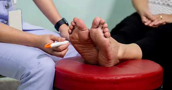
It is well known that diabetic foot ulcers are difficult to heal and some of this is due to delayed or abnormal fibroblast activity and quality. It has been shown that fibroblasts from chronic wounds (such as diabetic ulcers) display abnormalities e.g., decreased proliferation, altered patterns of cytokine release, early senescence and abnormal metalloproteinase activity. As a result, allogenic fibroblasts grafted to a bi-layered skin substitute have been used to support local dysfunctional fibroblasts, however, these do not persist indefinitely but do serve as a source of growth factors and cytokines. There is some evidence that electrical stimulation (ES) in non-diabetes-related skin can increase the growth of fibroblasts.
Furthermore, fibroblasts exposed to ES have also been shown to secrete higher levels of cytokines and growth factors, compared to non-exposed cells. This study evaluated the effect of ES on diabetic human skin fibroblasts (DHSF). The investigators examined DHSF adhesion, growth, the secretion of cytokines and growth factors compared with an experimental control group. The control group were exactly the same as the test group but without ES.
The authors also investigated the long-term effects of ES on DHSF shape and growth. The DHSF were harvested and isolated from skin tissues collected from patients with diabetes (61 to 80 years old) following leg amputation surgery. These were seeded (5 × 10⁴ cells/cm²) on the membranes in the electrical cell culture device and cultured for 24 hours at 37˚C in a 5% CO2 humid atmosphere. ES at various intensities of 100, 80, 60, 40 and 20 mV/mm were then applied or not to the cells for 6 or 24 hours. Following each stimulation period, the culture medium was refreshed and the cells were cultured for an additional 48 hours. The effect of ES on fibroblast adhesion and growth was evaluated using Hoechst staining, MTT and trypan blue exclusion assays. The secretion of cytokine and growth factor was assessed by cytokine array and ELISA assay.
This study reports that ES at 20 and 40 mV/mm promoted DHSF cell adhesion, viability and growth. It also demonstrated that ES decreased the secretion of pro-inflammatory cytokines IL-6 and IL-8 yet promoted growth factor FGF7 secretion during 48 hours after ES. Additionally, it also showed that the effect of ES DHSF growth was maintained up to 5 days after ES.
This paper is not the easiest to read but the study design is good and it is worth reading. As always, nothing replaces the fundamental triad of diabetic ulcer healing — in/outflow, infection control and offloading — but ES may be a worthwhile adjunct therapy.