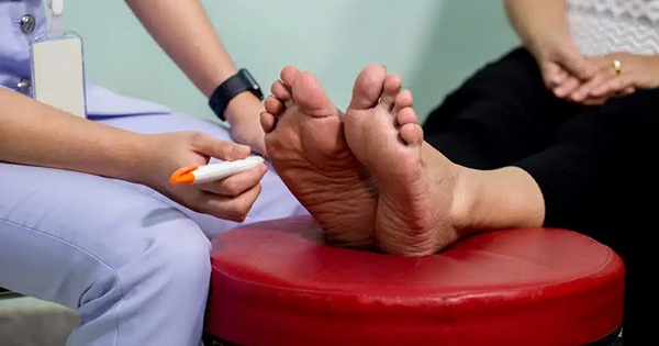
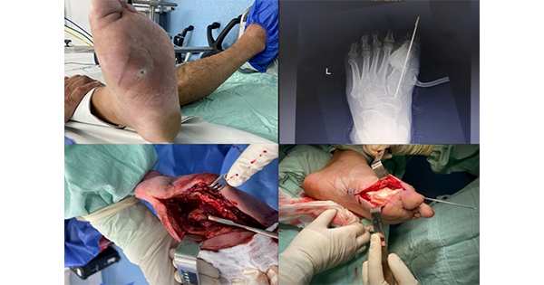
It has been estimated that at some stage in their lives, 10% of people with diabetes will develop a diabetic foot ulcer (DFU) (National Institute for Health and Clinical Excellence [NICE], 2015). According to NICE, foot ulcers are evident in over 80% of amputations in patients with diabetes. Within 5 years of amputation, up to 70% of amputees die, and approximately 50% die within 5 years of DFU onset (NICE, 2015). DFUs therefore have a significant impact on morbidity and mortality. For these reasons, it is important that DFUs are treated promptly and appropriately to optimise healing conditions and minimise impact on patients’ quality of life. Optimum management will also benefit health-care providers, as the treatment of DFUs is associated with considerable clinical and financial costs in the UK and elsewhere.
The financial impact of foot ulcers
The EURODIALE study, which used European prevalence data, estimated that DFU care costs up to 10 billion Euros, or £8.9 billion, per year (Prompers et al, 2008). DFUs, therefore, have a considerable financial impact on the NHS by way of costs generated in primary care, prolonged hospital stays, outpatient costs and community care. A 2012 report by NHS Diabetes estimates that approximately £650 million is spent each year on the treatment of foot ulcers or amputations (NICE, 2015). In another report, the total expenditure on DFU-related healthcare in 2014–15 in England was estimated to be £1 billion (Kerr, 2017). A US study published in 2014 with the objective of estimating the yearly, per-patient incremental burden of DFUs concluded that ‘DFU imposes substantial burden on public and private payers, ranging from $9–13 billion in addition to the costs associated with diabetes itself’ (Rice et al, 2014).
Minimising wound disturbance
Although dressings form an essential element of wound management, dressing-associated complications may hinder wound-healing progression and cause unnecessary distress to patients. Potential disturbances to wounds can result from suboptimal dressing choice. There are multiple ways in which a wound dressing that is in close contact with the wound bed and surrounding skin can disturb or damage the wound. These include (Rippon et al, 2012; Messaoud et al, 2018):
- Sub-optimal temperature
- Chemical imbalances
- Chemical stress
- Sub-optimal moisture balance
- Adherence
- Mechanical stress
- The presence of foreign bodies.
In recent years, the literature has focussed on trauma and pain caused by the repeated application and removal of dressings that adhere to the wound bed (World Union of Wound Healing Societies, 2004), as this damages the fragile wound or periwound skin and can result in considerable suffering for patients (Rippon et al, 2012). Ultimately, such trauma can lead to an increase in wound size, exacerbate pain and delay wound healing (World Union of Wound Healing Societies, 2004).
Although optimal dressing choice is important in achieving good healing progression, it also has a role in minimising the frequency of dressing changes, so allowing healing to progress uninterrupted. The frequent removal and reapplication of dressings can delay healing via mechanical disturbance of the wound-healing process, temperature loss at the wound site (affecting the cellular healing process) and a potential increase in the influx of harmful bacteria to the wound site (Rippon et al, 2015). Wound healing may be hindered further due to psychological stress and pain during dressing changes (Rippon et al, 2015).
In addition to optimising the frequency of dressing changes, dressings should be selected that manage the volume of exudate present, conform well to the wound, are comfortable to wear, are easy to use, minimise unnecessary wound disturbance and are cost-effective (Chadwick and McCardle, 2015).
Exudate management
A dressing’s ability to absorb and retain wound exudate is a key factor influencing wear time (Rippon et al, 2015). Although exudate formation is a normal part of the wound-healing process and an essential component of healing, excessive exudate that is not managed effectively can have a negative impact on the patient (Tickle, 2016).
Exudate is associated with a number of complications, including:
- Leakage
- Consequent maceration
- Malodour
- Pain and discomfort
- Psychological and psychosocial problems.
All of these complications can be detrimental to a patient’s quality of life (Faucher et al, 2012; Benbow, 2015; Moore and Strapp, 2015; Rafter et al, 2015; Tickle, 2016). Chronic wounds, such as DFUs, may produce high levels of exudate as a result of a prolonged inflammatory response preventing progression to the next phase of the healing trajectory (Chadwick and McCardle, 2015).
The ideal wound dressing should have optimal fluid handling ability (absorption and retention of exudate and its components, even under pressure); limit leakage; limit the spread of exudate to the periwound area (thus reducing the risk of maceration); and act as a barrier to prevent bacterial ingress.
Optimising adhesion
The adhesion of dressings is a factor that can affect dressing wear time. The ability of a dressing to conform to body contours helps ensure optimal adhesion (Rippon et al, 2015). However, according to International Best Practice Guidelines (2013), many dressings are not specifically designed for use on the foot and are consequently hard to apply over or between the toes and around the curvature of the heel. Additionally, there are no best practice guidelines to aid in selection of the most suitable dressings for such awkward sites.
Mepilex® Border Comfort
Mepilex® Border Comfort, which is marketed outside of the UK as Mepilex® Border Flex, is an all-in-one self-adherent soft silicone coated foam dressing (Figure 1). This dressing is designed for use on a wide range of exuding wounds, such as pressure ulcers, leg and foot ulcers, traumatic wounds (e.g. skin tears) and surgical wounds. It can also be used on dry/necrotic wounds in combination with gels. It comprises:
- A wound contact layer consisting of soft silicone adhesive (Safetac®) and a film carrier
- A flexible absorbent pad consisting of three layers: an absorbent foam, a non-woven spreading layer and a retention layer with superabsorbent fibres (the wound pad is partly perforated with Flex™ cut technology)
- An outer film that is breathable but impermeable to water, providing a barrier to external contaminants.
Dressings containing Safetac® wound contact layers readily adhere to intact dry skin and will remain in situ on the surface of a moist wound or damaged surrounding skin without adhering to these fragile tissues (White, 2005). Consequently, such dressings can be applied and reapplied without causing damage to the wound or stripping the epidermis in the periwound region (Meaume et al, 2003. They also minimise pain during dressing removal (Woo et al, 2009; Patton et al, 2013). The gentle but effective seal that forms between the intact skin and a dressing with Safetac® inhibits the movement of exudate from the wound onto the surrounding skin, thereby helping prevent maceration of the periwound region (White, 2005).
Flex™ technology makes Y-shaped cuts in the retention and spreading layers of the absorbent pad. These cuts contribute to the flexibility and conformability of the dressing and help prevent early detachment (Mölnlycke Health Care, data on file).
As well as being waterproof, thereby allowing patients to shower with the dressing in place, the backing layer of Mepilex® Border Comfort incorporates the unique Exudate Progress Monitor. This dot pattern allows for the easy tracking and recording of fluid as it spreads.
Case series
The following case studies were undertaken to evaluate the performance of Mepilex® Border Comfort when used in the management of DFUs. In each case, dressing changes were performed according to local clinical practice or when the dressing became saturated and at every follow-up visit to the podiatry clinic. Wound size and progression to healing were assessed at each visit to the clinic.
Case study 1
A 73-year-old male presented with a neuropathic DFU on the apex of the right first toe. He had a history of type 2 diabetes and chronic obstructive pulmonary disease. His osteomyelitis was being treated with clindamycin and ciprofloxacin.
The patient’s DFU had been present for 5 months and covered an area 109 mm. The wound bed was composed of 80% granulation tissue, 19% slough and 1% exposed bone (Figure 2a). Signs of local infection were present, including increased warmth and moderate levels of serosanguinous exudate. The periwound skin was healthy and intact. Before the use of Mepilex® Border Comfort, the DFU had been treated with UrgoTul® Absorb Border and offloading provided by a surgical shoe with a total contact insole.
A total of 11 Mepilex® Border Comfort dressings were used over a 25-day period. The patient did not experience pain at dressing change during this time. There were no clinical signs of local wound infection and the periwound skin remained healthy and intact throughout.
At the initial follow-up visit, the condition of the wound bed had improved slightly and consisted of 85% granulation tissue; however it remained unchanged after this time. There was a slight decrease in the volume of exudate by the third visit. The wound steadily decreased in size during follow-up period (Figure 2a–c). On day 25, the wound measured 62.5 mm2 and was 43% smaller than on day 1. Despite the reduction in size, the wound depth remained unchanged.
Case study 2
A 54-year-old male presented at the clinic with an ulcer at the amputation site of his right fifth toe that had penetrated to the bone. He had a history of type 2 diabetes, peripheral vascular disease and neuropathy. At the time of presentation, he was taking co-amoxiclav for osteomyelitis.
The 4 mm2 DFU (Figure 3a), had been present for 10 months. The wound was a deep sinus. There were no clinical signs of infection. Moderate levels of clear/serous exudate were observed and the periwound skin exhibited slight maceration. The wound had previously been treated with either Melolin or Telfa™ dressings. A Dura sandal with total contact insole off-loaded the wound.
Mepilex® Border Comfort was applied (Figure 3b), and the progress of treatment monitored over a 27-day period, during which the patient attended four follow-up visits.
The wound area at the final follow-up visit was 2 mm2. The wound depth steadily decreased from 2 mm on day 1 to 1.5 mm on day 27. At the second visit (Figure 3c), the periwound skin was healthy and intact. Due to the unavailability of Mepilex® Border Comfort between the second and third visits, however, the patient used a Telfa™ dressing secured with tape at this time, which macerated the periwound skin. Mepilex® Border Comfort use was resumed on the third visit and at the final follow-up the periwound skin was again healthy and intact, see Figure 3d. The frequency of dressing changes was reduced from daily with Telfa™ and Melolin to three times a week with Mepilex® Border Comfort.
The volume of exudate remained moderate throughout the 27-day period. It did, however, change from being serous/creamy yellow to bloody in nature after the second visit.
At baseline, the patient reported pain on dressing change to be 4 out of 10 on a visual analogue scale ranging from 0 (no pain) to 10 (maximum pain ever). From the third follow-up visit onwards he experienced no pain at dressing change. The patient stated that “The ulcerated area has become less painful” and “the aching in the wound area at night is reduced” with the use of Mepilex® Border Comfort.
Case study 3
A 59-year-old male presented at the clinic with a 5-month history of DFU located across the left, fourth and fifth toe amputation site. The patient had type 2 diabetes, peripheral vascular disease, neuropathy and osteomyelitis. The osteomyelitis was being treated with intravenous antibiotics. The patient had recently undergone an angioplasty procedure.
The DFU had an area of 208 mm2 and a depth of 2 mm, and was producing a moderate amount of serous exudate. The wound bed was composed of granulation tissue with a small amount of slough, and the periwound skin was healthy and intact, (Figure 4a). The wound had previously been treated with Aquacel®, Allevyn and Flaminal® Forte. A walker boot had been provided for offloading.
Treatment with Mepilex® Border Comfort was monitored over a 29-day period, during which the patient attended three follow-up visits at the podiatry clinic. Nine dressings were used during the study period and the patient reported no pain during dressing change procedures.
The wound bed contained granulation tissue at the first follow up. After 2 weeks of treatment, the central area had healed (Figure 4b), resulting in two individual wounds measuring 10 mm × 4 mm × 2 mm (wound 1) and 10 mm × 6 mm × 2 mm (wound 2). The amount of exudate was low from this point onwards.
The wounds continued to decrease in size throughout the period of study, with wounds 1 and 2 decreasing by 37.5% and 90%, respectively. At the final follow-up visit, however, despite the significant reduction in size, the depth of each wound had increased (wound 1 to the bone, and wound 2 to 4 mm) (Figure 4d). At this time, the periwound skin was healthy. The bed of wound 1 was predominantly granulation tissue, but the bed of wound 2 remained unchanged.
Podiatrists’ assessment of the dressing
In all three cases, the podiatrists on average rated the product as ‘Excellent’ in terms of its handling ability, ease of application, adherence, ability to allow wound inspection, handling of exudate and ease of removal. They observed that it conformed well to the wounds (Figure 2d, 3b and 4c), which were in difficult-to-dress areas. In case 3, they reported that the ability of Mepilex® Border Comfort to remain in place and its successful management of exudate had helped to reduce the number of dressing changes required.
Discussion
There were marked reductions in wound area in all three case studies following the use of Mepilex® Border Comfort, indicating good progression to healing.
Patient responses indicate that the use of Mepilex® Border Comfort is not generally associated with pain during dressing change. Feedback from patient 2 indicates that the use of this dressing is also associated with an absence of pain between dressing changes and reduced nocturnal pain, which in this individual was thought to be due to neuropathy.
Pain and stress are associated with reduced rates of healing (World Union of Wound Healing Societies, 2004; Rippon et al, 2015), therefore formulations that reduce pain and any associated anticipative stress at the time of dressing change potentially optimise the chances of healing. This may be reflected in the marked reductions in wound area seen when replacing previous dressings with Mepilex® Border Comfort in this case series.
Podiatrists indicated that Mepilex® Border Comfort handled exudate well, and therefore minimised maceration risk, that it was easy to apply and adhered well. These factors may have contributed to a reduction in the number of dressing changes required. The reduced frequency of dressing changes with Mepilex® Border Comfort compared to prior dressings is likely to have allowed healing of these moderately exuding wounds to progress.
The ease of application at the wound site and conformability of Mepilex® Border Comfort are of particular relevance to DFUs. The ulcers in this case series were located in awkward areas of the anatomy, making other types of dressing prone to dislodge. Dislodged dressings lead to reduced protection, reduced patient comfort, an increased risk of unnecessary disturbance to the wound, possible exposure to pain and stress during dressing change, and increased risk of contamination. Additional dressing changes due to poor adherence are associated with increased costs associated with nursing time, materials, pain medication and medication for dressing-related trauma.
The results of this case series thus suggest that Mepilex® Border Comfort optimises the wound healing environment by managing exudate and adhering well to the foot, eliminating unnecessary dressing changes, while being non-traumatic during removal and comfortable while in place.


