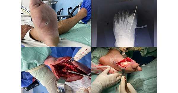
The prevalence of diabetic foot ulcers (DFUs) varies across different regions, with rates ranging from 1.7% in the UK to 12% in eastern Indonesia. Research findings highlight the importance of understanding and addressing regional disparities in DFU prevalence to improve patient outcomes and healthcare management strategies (Boulton et al, 2005; Yusuf et al, 2016).
The progression of infection in DFUs leading to bone involvement triggers an increased influx of inflammatory cells, which can be quantified by measuring C-reactive protein (CRP) (Lipsky et al, 2020). CRP is a marker synthesised in the liver during acute and chronic inflammatory conditions, falling under the category of acute-phase reactants. The elevation of CRP levels at varying rates reflects the severity of illness correlated with the extent of the inflammatory response orchestrated by inflammatory cells and cytokines (Markanday, 2015). Therefore, assessing the level of CRP is imperative in diagnosing the severity of DFUs and predicting their healing trajectory for effective wound management strategies.
Recent research has extensively examined the use of CRP in diagnosing the severity of DFUs. Umapathy et al (2018) found a significant correlation between circulating CRP levels and the severity of DFUs. Similarly, Fleischer et al (2009) study on diabetic foot osteomyelitis (DFO) revealed that CRP exhibited high sensitivity in diagnosing osteomyelitis as a standalone marker. Additionally, Jeandrot et al (2008) demonstrated that CRP at a cut-off value of 17 mg/dL displayed superior sensitivity and specificity in distinguishing between infected and non-infected DFUs.
The present literature review highlights a critical gap in the current understanding of CRP as a diagnostic tool for DFUs and DFO. While several studies have investigated the utility of CRP in diagnosing grade 2 DFUs and DFO, inconsistencies in the reported findings suggest a need for further research. Specifically, the mean cut-off value, sensitivity, specificity, and area under the curve associated with CRP in differentiating non-infected DFU from infected DFU without osteomyelitis were reported as 225.1mg/L, 77.4%, 84.3%, and 0.893, respectively. However, the diagnostic accuracy of CRP for DFO remains inconclusive due to a lack of consensus on appropriate cut-off values and significant variations in reported sensitivity and specificity values across studies. This underscores the need for future research to establish standardised guidelines for utilising CRP as a diagnostic marker in diabetic foot complications (Sharma et al, 2022).
The existing body of literature has elucidated the role of CRP in distinguishing between infection and non-infection in patients with DFUs and DFO. However, a notable gap in research pertains to the utility of CRP in differentiating between healing and non-healing DFU cases. As such, this study seeks to address this knowledge gap by examining the effectiveness of CRP as a biomarker for assessing the healing status of DFUs.
Methods
Study design and setting
The present investigation encompassed a prospective observational approach, wherein patients were meticulously monitored until the resolution or deterioration of their respective wounds necessitated amputation. The study was carried out at a specialised wound care facility situated in Indonesia, spanning from July 2022 to August 2023.
Participants
The research design of this study involved a specific set of inclusion and exclusion criteria, wherein patients aged 21 years and older with a confirmed diagnosis of type 2 diabetes were eligible for participation based on the 2024 American Diabetes Association [ADA] guidelines (ADA, 2024). These guidelines stipulated the presence of classic hyperglycaemia symptoms and a random plasma glucose level exceeding ≥200mg/dl (11.1mmol/l) as diagnostic criteria. Conversely, individuals presenting with wounds extending beyond the ankle were excluded from the study.
Data measurements: patient and wound characteristics
The study meticulously gathered patients’ demographic information, encompassing variables, such as age, sex, occupation, treatment of diabetes, wound aetiology, wound characteristics, anatomical site of the wound, severity classification based on the Wagner scale, presence of neuropathy and hypertension.
Laboratory parameters
Upon admission, patients underwent blood sampling prior to the commencement of antimicrobial therapy to analyse levels of haemoglobin, albumin and CRP. The quantification of these biomarkers was carried out by the clinic’s biochemistry laboratory.
Ethical consideration
The present research was carried out under the auspices of the Ethics Committee at the esteemed School of Health YARSI Pontianak, Indonesia (Ref. 028/KEPK/STIKes.YSI/X/2022). Prior to their participation, individuals and their family members were provided with a comprehensive explanation of the study’s objectives and procedures. Furthermore, participants were assured of their autonomy to discontinue involvement in the research at any point without being obligated to provide a rationale for their decision.
Statistical analysis
The study utilised descriptive statistics in the form of n (%) and median interquartile range (IQR) to present data. A comparative analysis between healed and non-healed was carried out employing statistical tests, such as the chi-squared test or Fisher’s exact test for categorical variables, and the Mann–Whitney U test for continuous variables.
In order to ascertain the likelihood of healing in DFUs, a linear regression analysis was employed with a significance level set at P<0.05. Prior to inclusion in the regression model, multicollinearity among the candidate variables was carefully evaluated to ensure the accuracy and reliability of the predictive model. The statistical analyses in this study were conducted utilising the Statistical Package for the Social Sciences (SPSS) version 26, produced by SPSS, Inc.
Results
The study included a total of 71 patients after excluding 28 individuals with incomplete data related to laboratory tests. Table 1 displayed no significant differences in age, gender, occupation, treatment of diabetes, trigger, wound status, wound location and hypertension (P>0.05). However, there were significant variations in the Wagner scale and neuropathy between the healed and non-healed groups (P=0.01 and P=0.04, respectively).
The results of the study revealed significant differences in haemoglobin, albumin, and CRP levels between the healed and non-healed groups in Table 2 of related laboratory tests (P=0.008, P=0.001, and P=0.001, respectively). Upon conducting multicollinearity testing with all candidate variables, such as the Wagner scale, neuropathy, haemoglobin, albumin and CRP, only CRP, Wagner scale, and neuropathy were found to be suitable for inclusion in the regression analysis. Subsequently, regression analysis (Table 3) identified CRP, Wagner scale and neuropathy as significant predictors of healing in diabetic foot ulcers, with P-values <0.001 for CRP and Wagner scale, and a P-value of 0.008 for neuropathy.
Discussion
This research endeavour delved into investigating the intricate interplay between CRP levels and healing outcomes in individuals affected with DFUs. After a thorough examination of 71 DFU patients in a prospective manner, our analysis unveiled a notable correlation between CRP levels and the eventual healing status of DFUs. The ramifications of DFUs, as a formidable complication in people with diabetes, are underscored by prolonged hospitalisation and escalating healthcare expenditures. As biomarkers such as CRP are progressively scrutinised for their prognostic value and capacity to gauge the severity of DFUs, this study contributes to the expanding body of knowledge on enhancing clinical management strategies for this challenging condition.
CRP is a crucial biomarker that indicates systemic inflammation and immune response in the body, commonly increasing in various inflammatory and infectious states. With both pro-inflammatory and anti-inflammatory characteristics, CRP plays a vital role in recognising and clearing foreign pathogens, as well as damaged cells by binding to specific molecules, such as phosphocholine, phospholipids, histones, chromatin and fibronectin. Furthermore, CRP can activate the classical complement pathway and stimulate phagocytic cells through Fc receptors to enhance the removal of cellular debris, damaged cells, apoptotic cells, and foreign pathogens (Nehring et al, 2023).
The findings presented in this study suggest a significant association between elevated CRP levels and non-healed DFUs. Previous research, including the study by Hadavand et al (2019), has consistently demonstrated a negative impact of high CRP levels on the healing process of DFUs. Additionally, meta-analyses, such as that conducted by Zhang et al (2022), further support the notion that CRP levels play a crucial role in predicting outcomes of DFU healing. These results underscore the importance of monitoring CRP levels in patients with DFUs to optimise treatment strategies and improve clinical outcomes.
The association between elevated CRP levels and infected DFUs has been extensively studied in the medical literature. Jeandrot et al (2008) underscore the significance of CRP as an early marker for identifying infections in DFUs. Moreover, persistently high CRP levels despite treatment may indicate a poor prognosis, prompting healthcare providers to consider more aggressive therapeutic interventions and enhance wound care practices. Conversely, a decrease in CRP levels is often indicative of improved healing outcomes and reduced ulcer severity, highlighting the clinical utility of monitoring this inflammatory marker in the management of DFUs.
CRP serves as a valuable risk parameter for identifying potential emergencies stemming from infections and inflammation in wounds, such as DFU. By prioritising preventative measures in emergency management, early assessment of CRP levels can offer a crucial early warning signal of impending emergencies. Additionally, monitoring CRP levels facilitates effective infection management by indicating fluctuations in the risk level of emergencies precipitated by infections.
Various screening tools have been proposed in the medical literature to detect sepsis or predict sepsis-related hospital mortality, yet none have demonstrated absolute accuracy. Despite this limitation, the utilisation of these tools is essential for facilitating early recognition and management of sepsis. It should be integrated into hospitals’ sepsis performance improvement initiatives, which have been linked to decreased mortality rates. The 2021 Surviving Sepsis Guidelines advised against using qSOFA in isolation for sepsis screening, favouring instead the use of SIRS, NEWS, or MEWS. Notably, none of these screening tools incorporates CRP levels as a component within their parameters (Evans et al, 2021; Kamath et al, 2023).
The association between the Wagner scale and wound healing outcomes in patients with DFUs has been extensively documented in previous studies. Recent research by Yang et al (2023) demonstrated a significant correlation between Wagner grade and prognosis in DFU patients with foot infections, with higher Wagner grades indicating a poorer prognosis. Lee et al (2024) further elucidated that neuropathy and aberrant cellular and inflammatory processes play a crucial role in hindering the healing process and increasing susceptibility to infection in diabetic foot ulcers.
The utilisation of laboratory examination of CRP levels during the initial assessment and subsequent monitoring within wound care programmes serves as a valuable tool in early detection of infections, thus aiding in the prevention of emergent complications and ultimately enhancing patient outcomes. By integrating CRP levels alongside clinical assessments, healthcare providers can improve diagnostic precision for DFU infections, track treatment efficacy and assess alterations in infection-related risk levels. This comprehensive approach to wound care management underscores the importance of utilising CRP levels as a crucial component in ensuring timely and effective interventions for patients with DFUs.
It is imperative to acknowledge the limitations of this study, particularly in its single-centre design, which may constrain the applicability of the results to a broader population. Moving forward, there is a pressing need for multicentre studies to validate these findings and delve deeper into the relationship between CRP levels and the healing of DFUs.
Conclusion
In light of the aforementioned findings, it is conceivable that CRP levels may serve as a prognostic indicator for the healing of DFUs within controlled laboratory environments, with potential implications for clinical practice.


