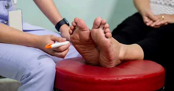
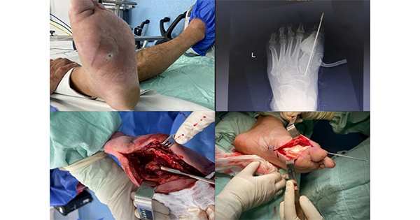
There is an average of 59,000 active foot ulcerations at any one time in England and there are about 135 amputations of the foot every week (Diabetes UK, 2016). Posnett and Franks (2008) estimated that around 200,000 people in the UK have a chronic wound at any one time. They also stated that in 2008, it was estimated that chronic wounds cost the NHS about £4bn a year. Given the extent of this problem, wound care needs to be both clinically effective and cost effective to help chronic wounds, such as diabetic foot ulcers, heal before they have such a devastating effect (Vowden, 2011).
Topical antimicrobial wound care products have become increasingly important in the treatment of infected wounds as there has been a necessary move away from the widespread use of systemic antibiotics due to global antibiotic resistance increasing (World Health Organization [WHO], 2014). Alongside this, the increased focus on biofilms and the role they play in chronicity has led to a change in approach for the care of chronic wounds to ensure that a variety of methods are used to disrupt biofilms.
This article will present the current thinking on biofilms, discuss how they are tackled when treating chronic wounds and diabetic foot ulcers, and will present a case study of a wound cleansing product that is able to disrupt biofilms and encourage a normal wound healing trajectory in previously hard-to-heal wounds.
What is a biofilm?
Percival et al (2000) described biofilm as “a community of microorganisms, either evident as mono-species or mixed species of microorganisms, attached to the surface (abiotic or biotic) or each other, encased within a matrix of extracellular polymeric substances and internally regulated by the inherent population”.
A biofilm forms a complex microbial community that is encapsulated in an extracellular polysaccharide matrix (glycocalyx). The glycocalyx is composed of proteins, polysaccharides and extracellular DNA.
The matrix of sugar and protein shields the microbial contents against the effects of the immune system of the host, as well as from many topical and systemic antimicrobial agents. The organisms within the biofilm cannot be detected using a normal wound culture method (Keast et al, 2014) and the biofilm acts as a collective entity, and is stronger and more resistant than its individual parts.
Single-celled organisms generally exhibit two distinct modes of behaviour:
- Planktonic: a free-floating form in which single cells float or swim independently in a liquid medium
- Sessile: an attached state in which cells are closely packed and firmly attached to each other, usually on a solid surface.
A change in behaviour in the single-celled organisms is triggered by many factors, including quorum sensing, as well as other mechanisms that vary between species (International Wound Infection Institution [IWII], 2009).
The biofilm phenotype predominates in nature (Costerton, 1995) and biofilms constitute the majority of bacteria in pathogenic ecosystems (Costerton et al, 1999). Harmful biofilms have been found attached to non-biological surfaces and medical devices, including artificial hips, heart valves, catheters, intrauterine devices, and an array of other medical prostheses. They are also found in the wound bed of chronic wounds.
Biofilms play a significant role in the inability of chronic wounds to heal. It is estimated that over 90% of chronic wounds contain bacteria and fungi living within a biofilm. Each aggregation of microbes creates a distinct biofilm with differing characteristics so that a clinical approach has to be tailored to the specifics of a given biofilm (Attinger and Wolcott, 2012).
How do biofilms form?
Stage one: reversible surface attachment
Free-floating, planktonic bacteria attach to surfaces and gradually form biofilms. Initially, the attachment is reversible.
Stage two: permanent surface attachment
The bacteria multiply and become sessile as the attachment becomes more permanent. They communicate with each other using quorum sensing and change their gene expression patterns in order to become stronger and survive.
Stage three: slimy protective matrix/biofilm
After the attachment has become more permanent the bacteria secrete a protective matrix known as extracellular polymeric substance (EPS). This is the initial biofilm (Phillips et al, 2010).
The composition of EPS varies according to the microorganisms present, but generally consists of polysaccharides, proteins, glycolipids and bacterial DNA. The DNA released by living or dead bacteria is thought to provide an important structural component of the EPS. Secreted proteins and enzymes help the biofilm become firmly attached to the wound bed. Fully mature biofilms continuously shed planktonic bacteria, microcolonies and fragments of biofilm, which can disperse and attach to other parts of the wound bed or to other wounds, forming new biofilm colonies (Wolcott et al, 2008).
How fast can biofilms form?
Planktonic bacteria can attach within minutes and become stronger and more tolerant to antibiotics, antiseptics and disinfectants, within 6–12 hours. A mature biofilm colony can develop within 2–4 days, depending on the species and growth conditions. Even after biofilms have been disrupted through debridement, a mature biofilm can reform within 24 hours (Philips et al, 2010).
The chronic inflammatory response is not always successful in removing the biofilm and it has been hypothesised that the response is in the interest of the biofilm. By inducing an ineffective inflammatory response, the biofilm protects the microorganisms it contains and increases exudate production, which is a source of nutrition that helps to perpetuate the biofilm (Phillips et al, 2010).
Biofilms employ multiple defence mechanisms, which lead to increased resistance to antibiotics, antiseptics and host immune defences. The ways that biofilms defend against immune responses are listed in Box 1.
How do biofilms delay wound healing?
The biofilm delays healing without causing obvious clinical infection. It evades the host’s natural defences and resists attacks from antibiotics and neutrophils.
Biofilms have a greater virulence than planktonic bacteria and are remarkably difficult to treat with antimicrobials. They can be up to 4,000x more resistant. The presence of a biofilm ensures that a chronic inflammatory state is maintained, which feeds the wound’s chronicity.
Treating chronic wounds
Microorganisms in a wound should be regarded as a collective ecosystem, or community, rather than as individual species.
The potential of biofilm maturation on the surface of dressings, principally gauze, may increase the release of biofilm virulence factors, such as acyl homoserine lactones, into the wound while also seeding the wound with planktonic bacteria, thereby contributing to the maintenance of a chronic inflammatory state (Rhoads et al, 2008).
The presence of ‘slime’ or slough on the dressing can be sufficient to suspect the presence of biofilm.
Difficulties encountered when treating biofilms
It is difficult to deliver antimicrobial agents through the EPS matrix at effective concentrations. Concentrations required to penetrate the biofilm are thousands of times greater than necessary to treat planktonic bacteria — sometimes higher than the toxicity threshold of the antibiotic.
The cells at the base of the biofilm operate at a very low metabolic rate. They ‘hibernate’, waiting for nutrients to revive them. The low metabolic rate of the sessile organisms drastically reduces the effectiveness of antibiotics and promotes antibiotic resistance (Hall-Stoodley and Stoodley, 2009).
How to manage a wound that has a biofilm infection
Treatment must suppress the biofilm, but not damage the host defences and/or the healing mechanisms. The treatment options are:
- Topical agents
- Broad spectrum antibiotics
- Debridement.
They should be used concurrently rather than consecutively (Wolcott et al, 2008).
Anti-biofilm agents include: honey which blocks lectin PA-IIL and Pseudomonas (P.) aeruginosa adhesion in vitro (Lerrer et al, 2007); silver, which has been shown to destabilise the biofilm matrix in vitro (Chaw et al, 2005); and iodine cadexomer, which soaks up Staphylococcus (S.) aureus cells, directly destroys biofilm structures, collapses glycocalyx during dehydration and can kill S. aureus cells within biofilms (Akinyama et al, 2004).
Removal of biofilms
Debridement
Partial removal of biofilm will lead to the expression of genes and increased virulence as
the biofilm adapts and become more active. Therefore, a comprehensive strategy to manage biofilm is required, and repeated debridement of the biofilm is required at least once every week (Wolcott et al, 2008).
Rapid removal of exudate from the wound bed may prevent the biofilm from making full use of the nutrients in the biofilm.
Wound cleansing
Wound cleansing is conducted to remove contaminants, debris, dressing remnants and superficial slough. It is possible to use tap water or sterile water, but scrubbing the wound can cause trauma. Antiseptics used for wound cleansing include:
- Alcohols
- Aldehydes
- Oxidative
- Phenols
- Quaternary ammonium compounds
- Guanidine.
The most widely available clinical antiseptics are guanidine (PHMB), chlorhexidine and octenidine dihydrochloride.
Octenisan
Octenisan® (schülke) is a hair and body wash that contains otenidine and allantoin, and has good skin-care and antimicrobial properties. It is suitable for all skin types — even skin that is sensitive to soap or susceptible to allergies — and for mild and gentle antimicrobial washing of patients prior to surgery, washing amputation stumps, and providing support for infection prevention and avoidance of relapses and secondary infections. Octenisan is epecially suitable for use on intensive care and infection wards. Octenisan wash mitts are also available, and can be used for cleaning the skin around the wound and are effective against a broad range of microorganisms (including multi-resistant strains).
Octenilin
Octenilin® (schülke) wound rinsing solution irrigates, cleanses and decontaminates chronic skin wounds. It can remove biofilms and has been shown to be able to reduce the number of pathogens in a wound and is more effective than Ringer’s solution or isotonic saline solutions commonly used in hospitals.
It is fast and easy and safe to use as standard practice. It contains ethylhexylglycerin, a surfactant that reduces the surface tension of a liquid so that the liquid can spread further. It also increases the wetting effect — loosening the biofilm’s devitalised tissue. It has a lower surface tension than Ringer’s solution. It can break down both immature and established biofilms.
Case studies
Case study one (GB)
The patient was a 75-year-old woman with diabetes and multiple diabetic ulceration of the foot. She had a partial forefoot amputation and, after 4 weeks of treatment with povidone-iodine, there had been no significant improvement and the wound had a dry, necrotic fibrinous coating (Figure 1).
After one week of treatment with octenidine-based solution and application of octenilin wound gel, it was easy to detach the crust and the wound bed was exhibiting granulation tissue (Figure 2). Two weeks later, there was clear progress in epithelialisation and further detachment of the fibrin coating (Figure 3). After 7 weeks of treatment wound closure was nearly complete (Figure 4).
The use of Octenilin was able to clean away debris, break up the biofilms that had formed and prevent them reforming by removing its nutrients, and breaking the cycle of chronicity and its use set the wound onto a normal wound healing trajectory.
Case study two (NR)
This case study focuses on a man admitted to the acute hospital for a non-wound related condition. Patient X was 52 years old and had a past medical history of untreated hepatitis C and presented to the hospital in an undernourished state. He also had a history of deep vein thrombosis in his right leg, 3 years previously. He was an intravenous drug user who was using two bags of heroin daily and had regularly used his right pretibial area as site of choice when injecting. During the admission process, he was found to have extensive ulceration to his right lower leg. This case study will look at the dressing regimen, while an inpatient and the concurrent use of an antimicrobial solution, Octenilin, and bacteria binding wound interface dressing, Cutimed® Sorbact® (BSN Medical) as a means of reducing the bioburden present in the wound and to encourage granulation and epithelisation.
On examination, he was found to have a 25 cm x 18 cm malodourous wound to his right lower leg, which spread from the anterior aspect laterally round to the posterior, with obvious signs of a biofilm being present (Figure 5). There was thick dark slough present with several areas of hard eschar spreading inwards from the wound edges and a purulent discharge was noted. Just above this large wound was the pretibial area where he had been injecting the drugs. This was an area that was a mixture of hard and soft eschars, and scar tissue. Several of the pretibial wounds showed evidence of pus in the wound beds and there were small bits of tissue embedded into the wounds as Patient X had been self-managing his wounds prior to admission without the aid of proper dressings for approximately one year. He had been treated with antibiotics on many occasions for infections in this area. Pain on dressing change was reported by the patient to be ‘excruciating’ and he, at one point, left the ward in order to use heroin as he found the pain unbearable. Arrangements were made for him to be prescribed a neuropathic analgesia as the pain he described appeared to be neuropathic in nature.
A biofilm occurs when bacteria multiply within a wound to a critical level and it can cause the wound to be stuck in the inflammatory response stage and reduces the possibility of the wound healing, thereby placing the wound into the chronic wound category. In order to enable the wound to move on to the proliferative stage, this biofilm needed to be removed and it was for this reason that the clinician used both the Octenilin solution and the Cutimed Sorbact.
Octenilin solution is an antimicrobial cleaning solution, which is marketed for cleansing the wound and removal of biofilm/slough/debris. Patients report that it is painless on application and this clinician has found it can have a definite effect on wound healing rates for many patients. By helping to lift slough and biofilm from the wound bed, it enables the application of other dressings that will help move the wound to the next stage. Cutimed Sorbact is a wound contact dressing that works by using hydrophobic interaction to permanently bind and remove bacteria from the wound bed without donating anything to the patient, thereby reducing the risk of product build-up and resistance.
Basic 32-ply gauze was soaked in Octenilin solution and then placed over the wound beds for two/three minutes. Each piece of gauze when removed was used to help lift the slough present on the wound bed. Cutimed Sorbact was then soaked in Octenilin solution and laid over the wound bed. A super absorber was used to manage the exudate between dressing changes and bandaging was applied. Removal of the Cutimed Sorbact was aided by covering the area with Octenilin solution to rehydrate it.
Ten days later at the fourth dressing change (Figure 6), the dark slough had been removed from the larger wound with an island of non-ulcerated skin visible, the wound edges are pink, and 10% granulation tissue is present with 90% slough. The pretibial area shows evidence of epithelisation occurring. A reduction in pain was reported and reduced exudate levels noted.
Three weeks after admission (Figure 7), there is healthy granulation tissue present to approximately 40% of wound bed with superficial yellow slough to 60% with underlying granulation tissue present. There is evidence of skin edges drawing in and both pain and exudate levels were greatly reduced. The pretibial area is showing healthy epithelisation tissue with a greatly reduced area of eschar formation. Patient X was able to tolerate wound bed cleansing post-Octenilin and gauze soak at this point.
Unfortunately, the clinician was unable to continue with Patient X’s dressings, due to a job change, and they are unaware if the dressing regimen they instigated was continued or indeed if the outcome was a healed ulcer.
Biofilm seems to be one of the favourite buzzwords within the tissue viability world at the moment and there are many products that claim to be able to reduce/remove this bioburden in order for wound healing to be able to take place. Both Octenilin solution and Cutimed Sorbact are quick and easy to use, and provide effective and reliable wound cleansing, as well as biofilm reduction.
While there is no formal evidence that using Octenilin and Cutimed Sorbact together reduces the healing time of wounds or reduces the bioburden of wounds quicker than other antimicrobials or individually, the clinician has encountered good results when combining these two dressing products. Reduced healing times, decreased pain levels and reduced bioburden loads have been achieved on many occasions when this particular dressing regimen has been prescribed.
Conclusion
The presence of biofilms in wounds is often the cause of chronicity. Biofilms allow planktonic bacteria to act like a multicellular organism with the ability to increase its defences and virulence. Octenilin irrigation solution and gel are fast and effective tools for wound management that support all other interventions that can be used to treat chronic wounds. Biofilms must be tackled using a range of concurrent methods, but disruption, debridement and cleansing is the key to removal and will help the chronic wound to heal.
Chronic wounds have a huge impact on quality of life and they are also extremely costly to treat. Diabetic foot ulcers that do not heal can worsen and lead to amputation or death. It is important that clinically effective treatments are used to help chronic wounds heal.


