
Best practice injection technique is critical to ensuring optimal glycaemic control. Those responsible for the care of people with diabetes must be aware of the dangers of fluctuating blood glucose levels, which are often a result of people injecting into areas of lipohypertrophy (Forum for Injection Technique, 2012). Factors within the injection process, such as the re-use of needles, failing to rotate injection sites and injecting into lipohypertrophic regions, can all contribute to the erratic absorption of an injectable therapy (Forum for Injection Technique, 2012). Although this may be understood in some professional circles, it may not be so obvious to those with diabetes using injectable therapies (De Coninck et al, 2010). This case study discusses how to detect lipohypertrophy and the consequences of failing to routinely examine injection sites.
Case study
This case study concerns a 63-year-old man who was diagnosed with type 1 diabetes in 1985, aged 35. His records were reviewed as a result of an unexplained hypoglycaemic episode while he was driving. Large areas of lipohypertrophy were subsequently detected.
It was recorded that he was involved in a road traffic incident relating to a hypoglycaemic episode in 1988. In 2013, a referral from the Yorkshire Ambulance Service was received following another road traffic incident related to hypoglycaemia in which significant injuries were sustained. When questioned, the individual could not offer a reason as to why he had been experiencing such episodes. He claimed that there were no lumps at his injection sites and that he regularly rotated them.
In 1991, he had a total daily dose (TDD) of 78 units. It was recorded at that time that he had overused abdominal sites and was advised to rest and rotate the sites regularly. Between November, 1989 and when he was seen in 1991 his HbA1c had been steady at around 75 mmol/mol (9.0%), but then fell to 55 mmol/mol (7.2%) after being asked to rest and rotate his injection sites.
In 1992, he was changed onto a multiple injection regimen with insulin isophane and insulin lispro with a TDD of 48 units. It was again documented that he needed to rest his abdominal area.
In 2003, it was documented that he was taking 72 units of insulin lispro per day and 24 units of insulin isophane, and he was using 8 mm needles. His job at the time involved driving 1000 miles a week and he reported that he deliberately ran his blood glucose high to avoid hypoglycaemia.
In November 2012, the Yorkshire Ambulance Service was required twice over a 3-day period for two hypoglycaemic episodes. Following these episodes, his diabetes team made contact with him. The hypoglycaemia had not been reported to the team by the patient, but through a local pathway. At this point, he reluctantly agreed to see a dietitian as it was clear he was not matching his carbohydrate intake to insulin therapy correctly. In March 2013, he reported that he had not had any further episodes of hypoglycaemia.
During a routine review of the man in August 2013, it was reported that his HbA1c had remained stable (66 mmol/mol [8.2%]) with no further episodes of hypoglycaemia. He reported on this visit to the clinic that he did not routinely change his insulin pen needles in the belief that he was “saving the NHS money”. It was therefore explained that needle reuse is detrimental to the good management of diabetes as it can cause leakage, clogging, trauma to the skin and lipohypertrophy, all of which can cause the suboptimal delivery and absorption of the insulin dosage (Vardar and Kizilci, 2007).
In December 2013, a further Yorkshire Ambulance Service referral was received following another hypoglycaemic episode. It became apparent that this had happened while the man had been driving home from work. The car was considered a “write off” and he sustained three broken ribs, although no other vehicles were involved. The incident resulted in his driving licence being suspended for three months, which had a significant impact on his daily routine.
When the diabetes team spoke to him on the phone, he reported that his blood glucose was 7 mmol/L at 18:20 when he planned to leave work. He was delayed leaving and did not leave until 20:00. The accident occurred shortly before 20:56, which is the time the ambulance was called. He had injected 22 units of insulin glargine in the morning at 06:00 and had not had any insulin during the day. He had eaten a banana and a chocolate bar at approximately 17:00 (with no insulin) and was not able to explain what caused the hypoglycaemia. Injection sites were discussed and he reported he had no lumps at his injection sites and that he rested and rotated them appropriately. Injection sites were discussed with the man and his wife on several occasions; his wife also reported that she was unaware of any lumpy areas.
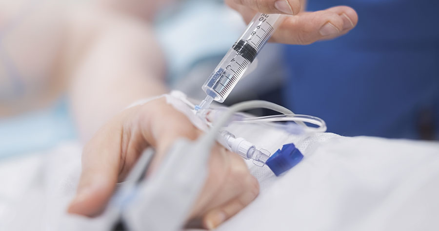
It was agreed he would phone again in a week with his blood glucose levels and, if possible, record them so that his team could accurately assess them. At this point, his blood glucose levels were mostly 7–13 mmol/L, but he had also had two further episodes of hypoglycaemia and again was unable to identify a reason. It was suggested that he have his injection sites physically examined by his diabetes team. The subsequent review revealed grossly overused injection sites in the inside thigh and abdomen (Figure 1).
Education was provided and he was advised that the inner thigh was not a recognised injection site and should not be used. He was advised that best practice injection technique requires him to rotate both the injection site and within the injection site, as recommended by the Forum for Injection Technique UK (2012). He was given 4 mm needles to replace the 5 mm needles he was currently using and was advised again about never reusing needles.
By February 2014, a period of regular contact with his diabetes team resulted in him accurately matching his insulin to his carbohydrate. His insulin glargine dose was reduced from 22 units to 16 units and his insulin dosage altered from up to 30 units, to no more than 12 units (working on a 1 unit/10g carbohydrate ratio). His maximum TDD has reduced from 112 units to 52 units per day. His HbA1c was essentially unchanged since his last visit despite over 50% reduction in TDD. His home blood glucose monitor readings were much more even. During June and July 2014, 70% of his blood glucose levels were in target, with only one reading of 3.8 mmol/L.
He still has the occasional hypoglycaemic episode but he understands why they occur. Further still, he recognises that his blood glucose levels are more even and predictable having followed the advice given about injection technique.
Lipohypertrophy: A problem for glycaemic control
As this case study shows, preventing lipohypertrophy with correct injection technique will contribute to good glycaemic control (Landau, 2012). Vardar and Kizilci (2007) found that injecting into lipohypertrophic areas can result in poor absorption and the need to inject higher doses of insulin. While the exact pathogenesis of lipohypertrophy is not fully understood, it has been suggested that the causative factors are likely to be linked to repeated insulin injections into the same small areas of tissue (Blanco et al, 2013).
It is possible that with each needle insertion and the resultant micro-trauma to the adipocyte cells (many tens of thousands obliterated with each needle insertion), the body activates wound healing, which includes hormones, chemicals and specialist cells flooding to the micro-trauma (Throw et al, 1990). This healing cascade takes place bathed in insulin-like growth factor-1 (Throw et al, 1990). Over time, repeated micro-trauma and wound healing in the presence of insulin appears to cause adipocyte cells to grow large and somewhat altered (hypertrophy) with large adipocyte cells mingled with smaller cells with evidence of fat droplets on the surface of adipocyte cells (Fujikura et al, 2005). Lipohypertrophic tissue is thought to be predominantly avascular, with constricted, reduced or possibly no blood vessels in some cases (Heinemann, 2010). Low levels of blood flow appear to alter the absorption rates of insulin, leading to increased doses being required when compared to healthy subcutaneous tissue (Heinemann, 2010).
Lipohypertrophy often develops due to repeated injection into the same area (Heinemann, 2010). Therefore, with the application of an injection site rotation system, it is less likely to develop. This is further corroborated within a recent study by Blanco et al (2013) that found that 98% of people with lipohypertrophy either did not rotate their injection sites, or rotated incorrectly, whereas only 5% of those who rotated correctly had lipohypertrophy. In addition, results showed that 49% of people with lipohypertrophy have glycaemic variation, compared with only 7% of people without lipohypertrophy, and 39% of people with lipohypertrophy have unexplained hypoglycaemia, compared to 6% of people without lipohypertrophy. This highlights the existence of lipohypertrophy as a significant concern when seeking to establish glycaemic control and reliable dosage delivery.
Discussion
The case reviewed involved a man who had attended a diabetes centre for many years. At the time of diagnosis he was taught carbohydrate counting, although his results suggest that he stopped practising this at some point until he was properly re-educated.
The management and care of injection sites is something that was documented as being discussed as far back as 1991, yet his sites were significantly overused and his wife also failed to appreciate that they were abnormal.
Over several years, he had a number of unexplained hypoglycaemic episodes. At this time, while injection sites were discussed, a physical examination of them does not seem to have been carried out. Since his road traffic accident, the diabetes team involved has undertaken physical examinations of injection sites. Another individual, who had a similar experience, reported:
“I hesitate to say it this early in case I jinx it, but it looks as if you have another success story with me and the injection site experiment. For hours on end yesterday I was 5 and 6 [mmol/L) and this was on vastly reduced insulin doses. I am astonished, literally, it feels like a miracle! Thank you so much for sharing the information with me and good luck for your ongoing important research.”
From my experience, nearly all individuals that have exhibited features of lipohypertrophy have denied having problems with their sites prior to the examination. Many inject into unusual sites, particularly the inner thigh and upper forearm. In addition, many are found to be unaware of, or had forgotten, the importance of correct injection technique. Some lesions were clearly visible and yet the individual had not brought these to the attention of the healthcare professional. It would seem that many people wrongly accept lipohypertrophy as a given consequence of using an injectable therapy and that many people are still re-using needles, increasing the risk of lipohypertrophy. This is often done in the misguided view that they are saving the NHS money.
As the case study highlights, the identification of lipohypertrophy and subsequent re-education can improve glycaemic control, and prevent or reduce the occurrence of hypoglycaemia. Follow-up education can also reduce the insulin dose required. These factors can result in a cost benefit to the healthcare economy, while improving the health and quality of life of the person with diabetes (Blanco et al, 2013).
Summary and best practice advice
As highlighted by the Royal College of Nursing (2012), many people are now using injectable therapies to achieve adequate glycaemic control and this can, in part, be attributed to people living with progressive type 2 diabetes for a longer period of time. The supervision and education delivered when injectable therapies are commenced may only be observed on one or two occasions. In my opinion, care of injection sites is vital in ensuring the on-going efficacy of injectable therapies. In everyday practice, diabetes teams rarely have the opportunity to look back over the history of specific individuals; however, repeated unexplained hypoglycaemic events act as a strong indication of lipohypertrophy, which can be rectified, or at least reduced, by reiterating information about best practice injection technique.
The Forum for Injection Technique (2012) has produced a useful quick-reference handheld tool that healthcare practitioners seeking to resolve unexplained glycaemic variation may find helpful. It suggests checking injection sites, injection technique, the type of insulin, injection equipment, food, activity and insulin dose. This type of resource acts as guidance and a checklist, which will help to ensure nothing gets overlooked. For reliable absorption, insulin should be injected into the subcutaneous layer; however, its thickness varies from site to site. Diabetes teams may see individuals benefit from using 4 mm pen needles as studies have found them to be appropriate for use by everyone, including obese people with diabetes (Laurent et al, 2007). It is known that 4 mm needles will consistently pass through the skin to the subcutaneous layer and reliably deposit insulin, as skin thickness is on average is 2 mm thick with little variation regardless of age, gender, BMI and ethnicity (Gibney et al, 2010).
Educational follow-up regarding injection technique is incredibly important as, although knowledge will be imparted at the initiation of a therapy, it is often at a later stage that problems relating to poor injection technique (such as lipohypertrophy) arise. It is, therefore, important to re-visit injection technique and examine injection sites as part of routine, ongoing management to continually achieve its optimal effect.
Conclusion
Healthcare professionals that come into contact with people with diabetes have a duty to thoroughly check an individual’s injection technique, educate where applicable, and to palpate for lipohypertrophy whenever possible. Where lipohypertrophy is identified, it is likely that insulin doses will need to be reduced to prevent hypoglycaemia. No irregularities should be ignored or underestimated and healthcare professionals must not rely on problems with injection sites being voluntarily brought to their attention.


The risk factors and what might be done to address them.
24 Mar 2025