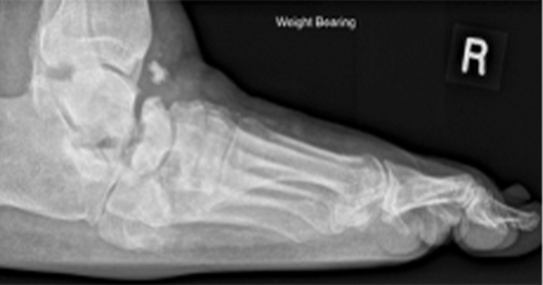
It has been estimated that an ulcer will develop on the foot or ankle of 15% of people with diabetes during their lifetime (Boulton, 2004). Various adjunctive physical therapeutic modalities, including ultrasound, electrotherapy and electromagnetic therapy, have been proposed for the treatment of lower-limb ulcers in people with diabetes (Cullum et al, 2001).
Photobiomodulation therapy is a technique whereby low-level polychromatic, coherent light is applied to injured dermis in an attempt to improve wound healing. The non-thermal effects of light between 1–10 J/cm2 on biological tissues has been shown to be beneficial in cell culture studies (Brondon et al, 2005).
Using National Aeronautics and Space Administration-developed light-emitting diode (LED) technology, Whelan et al (2001) reported in vitro increases in the growth of mouse fibroblasts, rat osteoblasts, rat skeletal muscle cells and human epithelial cells. Using the same light source, clinical reductions in wound size in rat studies, and reductions in healing time in human volunteers from the US Navy, were also reported (Whelan et al, 2001). However, data on the use of photobiomodulation therapy to treat diabetic foot ulcers are limited. In their review, Forney and Mauro (1999) concluded that further research is required to evaluate the role of lasers and photobiomodulation therapy in diabetic foot ulcer closure.
The retrospective cohort study reported here assessed the efficacy of photobiomodulation using LED (PLED) on the closure of diabetic foot ulcers.
Methods and participants
Twenty people who presented with diabetic foot ulcers between October 2000 and June 2002 at the Royal Preston Hospital were included in this study. Data were taken from podiatry and general medical records.
Participants were treated according to a standard protocol for diabetic foot ulcer management (Jeffcoate et al, 2004). This included regular sharp debridement of calluses surrounding the ulcer, use of walking or total-contact casts for off-loading and regular moist dressing. In addition to the traditional management protocols, participants received PLED therapy every 1–3 weeks until their ulcer healed, or until the end of the study period was reached (June 2002). Healing was defined as complete epithelialisation without discharge.
Foot care and PLED therapy were delivered by a trained podiatrist under the supervision of the clinic’s consultant physician. Ulcer size was taken as the diameter at its widest point. All participants underwent radiological assessment to exclude underlying osteomyelitis. Wagner’s scores (Wagner, 1981) were used to grade the severity of ulceration. Cellulitis was defined as an acute spreading infection extending at least 10 mm beyond the wound margin, with or without purulent discharge, and with or without evidence of systemic infection (e.g. fever, leucocytosis). Those diagnosed with cellulitis had wound swabs sent for bacterial culture and sensitivity testing and were treated with broad-spectrum antibiotics until the results of sensitivity testing allowed for narrowing to a more appropriate agent.
PLED therapy was delivered using the THOR-LX photobiomodulation unit (THOR Photomedicine, Chesham, UK; Figures 1–2). This unit incorporates two LED treatment probes: a single-point probe, and a cluster probe. The cluster probe comprised 34 × 660 nm 10 mW (power density 50 mW/cm2) and 35 × 950 nm 15 mW LEDs (power density 75 mW/cm2), with an average power density for the whole cluster probe of 62.5 mW/cm2. The single-point probe comprised 1 × 660 nm 10 mW LED (power density 50 mW/cm2) (THOR Photomedicine, 1998).
Treatment time was typically 60 seconds/cm2, with the cluster probe delivering 3.75 J/cm2 to each treatment site, followed by treatment with the single-point probe around the wound margin at 1 cm intervals. The probes were held as close to the wound as possible without touching its surface. Treatment with the appropriate probe continued until all of the wound, and the surrounding intact tissue, had received a complete dose.
Statistics
Data were analysed for computing odds ratio, 2-tailed Student’s t-test and Chi-squared test for P-values and Spearman’s Rank correlation test using SPSS software, version 11.5 (SPSS, Chicago, IL, USA). P-values <0.05 were considered to be statistically significant.
Results
Participant characteristics
Baseline participant characteristics are summarised in Table 1. Ulcers occurred most frequently in men aged 60–70 years (22.0%), and in women aged 80–90 years (15.0%). In this series, 25.9% of participants had type 1 diabetes, 66.7% had type 2 diabetes and 7.4% had diabetes secondary to chronic pancreatitis.
Of the 20 participants, nine (33.3%) had two ulcers and the rest (66.7%) had solitary ulcers. Analyses were conducted based on episodes of foot ulceration. A participant with more than one foot ulcer was considered “healed” only when all ulcers present during that episode of ulceration had healed. A total of 27 episodes of foot ulceration were treated during the study period.
Mean ulcer size was 15.7 ± 9.8 mm, taken at its widest point. Episodes of ulceration were most commonly limited to soft tissue (Wagner’s score 2; 77.8%), while 22.2% extended to bone (Wagner’s score 3). Ulcers were frequently located on the plantar aspect of the foot (92.6%), with the most common site of ulceration being the first metatarsal head (30.3%). Sixteen (59.3%) episodes of ulceration were painless. Seven (25.9%) episodes of ulceration were neuropathic, three (11.2%) were ischaemic, and 17 (62.9%) were neuro-ischaemic. There was a history of trauma in one (3.7%) episode.
Cellulitis was present in 33.3% of episodes of ulceration and resolved in an average of 1.9 ± 1.6 weeks. Wound swabs taken from ulcers with cellulitis revealed pathogenic microbes in 15 (55.6%) episodes of ulceration, the most common isolate being Staphylococcus aureus (48.3%).
Non-healing versus healed ulcers
Participant characteristics associated with those episodes of ulceration that healed, and those that did not, are summarised in Table 2.
In the cohort reported here, the main factors influencing healing were similar to those reported in previous studies investigating PLED therapy, namely peripheral neuropathy, arterial disease, poor vision, foot deformity and previous ulceration (Boyko et al, 1999; Abbott et al, 2002). Baseline ulcer size, described elsewhere as being a risk factor for non-closure (Oyibo et al, 2001; Sheehan et al, 2003; Zimny et al, 2004), was significantly larger in those that failed to heal, compared with those that healed (P=0.02). The other significant indicator of non-closure was cellulitis, which took significantly longer to resolve in those ulcers that failed to heal (P=0.04). Ulcer depth has similarly been reported to be a risk factor for non-closure (Oyibo et al, 2001; Treece et al, 2004), but was not found to be significantly different between the healing and non-healing groups in this cohort.
In a meta-analysis of ten randomised clinical trials of neuropathic ulcers receiving standard treatment, the closure incidence was 24.2% at 12 weeks and 30.9% at 20 weeks (Margolis et al, 1999), and Harrington et al (2000) found the overall incidence of closure for all diabetic foot ulcers to be 31%. In the cohort reported here, with its high overall incidence of lower-limb ischaemia (77.8%), the incidence of closure was 22.2% at 12 weeks, 29.6% at 20 weeks, and 48.1% by study end. These closure incidences, coupled with the fact that none of the participants underwent an amputation, suggest that PLED therapy improved wound healing in these diabetic foot ulcers.
Limitations
The limitations of this study are that it was not a randomised control trial, and that the number of participants was small. Larger studies investigating the use of PLED therapy for the treatment of diabetic foot ulcers are required.
Conclusion
Given the well-established association between ulcers, amputation and morbidity, any measure that aids closure of diabetic foot ulcers is positive.
PLED therapy is a safe, well-tolerated adjunct to standard wound care treatment for diabetic foot ulceration. While the efficacy of low-level phototherapy has yet to be confirmed by large randomised control trials, the results reported here suggest a positive role for PLED therapy in the promotion of healing of diabetic foot ulcers.
Acknowledgements
The authors would like to thank James Carroll of THOR Photomedicine for providing detailed technical specifications of the Thor LX photobiomodulation unit and for supplying photographs of the unit in use. The authors would also like to thank Professor Rayaz Malik of the Manchester Royal Infirmary for his advice on the preparation of this manuscript.


